4ETK | pdb_00004etk

Crystal Structure of E6A/L130D/A155H variant of de novo designed serine hydrolase, Northeast Structural Genomics Consortium (NESG) Target OR186


Experimental Data Snapshot

  • Method: X-RAY DIFFRACTION
  • Resolution: 2.70 Å
  • R-Value Free: 
    0.254 (Depositor), 0.254 (DCC) 
  • R-Value Work: 
    0.188 (Depositor), 0.190 (DCC) 
  • R-Value Observed: 
    0.188 (Depositor) 

Starting Model: experimental
View more details

wwPDB Validation 3D Report Full Report

Validation slider image for 4ETK

This is version 1.5 of the entry. See complete history

Literature

Design of activated serine-containing catalytic triads with atomic-level accuracy.

Rajagopalan, S.Wang, C.Yu, K.Kuzin, A.P.Richter, F.Lew, S.Miklos, A.E.Matthews, M.L.Seetharaman, J.Su, M.Hunt, J.F.Cravatt, B.F.Baker, D.

(2014) Nat Chem Biol 10: 386-391

  • DOI: https://doi.org/10.1038/nchembio.1498
  • Primary Citation Related Structures: 
    3TP4, 3V45, 4DRT, 4ESS, 4ETJ, 4ETK, 4F2V, 4JCA, 4JLL, 4JVV

  • PubMed Abstract: 

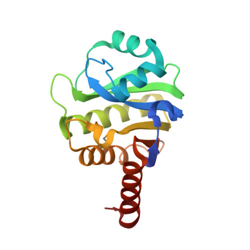
    A challenge in the computational design of enzymes is that multiple properties, including substrate binding, transition state stabilization and product release, must be simultaneously optimized, and this has limited the absolute activity of successful designs. Here, we focus on a single critical property of many enzymes: the nucleophilicity of an active site residue that initiates catalysis. We design proteins with idealized serine-containing catalytic triads and assess their nucleophilicity directly in native biological systems using activity-based organophosphate probes. Crystal structures of the most successful designs show unprecedented agreement with computational models, including extensive hydrogen bonding networks between the catalytic triad (or quartet) residues, and mutagenesis experiments demonstrate that these networks are critical for serine activation and organophosphate reactivity. Following optimization by yeast display, the designs react with organophosphate probes at rates comparable to natural serine hydrolases. Co-crystal structures with diisopropyl fluorophosphate bound to the serine nucleophile suggest that the designs could provide the basis for a new class of organophosphate capture agents.


  • Organizational Affiliation
    • 1] Department of Biochemistry, University of Washington, Seattle, Washington, United States. [2].

Macromolecule Content 

  • Total Structure Weight: 35.58 kDa 
  • Atom Count: 2,469 
  • Modeled Residue Count: 331 
  • Deposited Residue Count: 334 
  • Unique protein chains: 1

Macromolecules

Find similar proteins by:|  3D Structure
Entity ID: 1
MoleculeChains  Sequence LengthOrganismDetailsImage
De novo designed serine hydrolase
A, B
167synthetic constructMutation(s): 0 

Small Molecules

Ligands 1 Unique
IDChains Name / Formula / InChI Key2D Diagram3D Interactions
NA

Query on NA


Download:Ideal Coordinates CCD File
C [auth A]SODIUM ION
Na
FKNQFGJONOIPTF-UHFFFAOYSA-N
Modified Residues  1 Unique
IDChains TypeFormula2D DiagramParent
MSE
Query on MSE
A, B
L-PEPTIDE LINKINGC5 H11 N O2 SeMET

Experimental Data & Validation

Experimental Data

  • Method: X-RAY DIFFRACTION
  • Resolution: 2.70 Å
  • R-Value Free:  0.254 (Depositor), 0.254 (DCC) 
  • R-Value Work:  0.188 (Depositor), 0.190 (DCC) 
  • R-Value Observed: 0.188 (Depositor) 
Space Group: P 21 21 2
Unit Cell:
Length ( Å )Angle ( ˚ )
a = 78.196α = 90
b = 133.661β = 90
c = 29.937γ = 90
Software Package:
Software NamePurpose
PHENIXrefinement
PDB_EXTRACTdata extraction

Structure Validation

View Full Validation Report


Entry History 

Revision History  (Full details and data files)

  • Version 1.0: 2012-06-13
    Type: Initial release
  • Version 1.1: 2014-04-16
    Changes: Database references
  • Version 1.2: 2014-05-14
    Changes: Database references
  • Version 1.3: 2023-09-13
    Changes: Data collection, Database references, Derived calculations, Refinement description
  • Version 1.4: 2023-12-06
    Changes: Data collection
  • Version 1.5: 2024-11-06
    Changes: Structure summary