4EIS | pdb_00004eis

Structural basis for substrate targeting and catalysis by fungal polysaccharide monooxygenases (PMO-3)


Experimental Data Snapshot

  • Method: X-RAY DIFFRACTION
  • Resolution: 1.37 Å
  • R-Value Free: 
    0.149 (Depositor), 0.151 (DCC) 
  • R-Value Work: 
    0.116 (Depositor), 0.121 (DCC) 
  • R-Value Observed: 
    0.118 (Depositor) 

wwPDB Validation 3D Report Full Report

Validation slider image for 4EIS

This is version 2.1 of the entry. See complete history

Literature

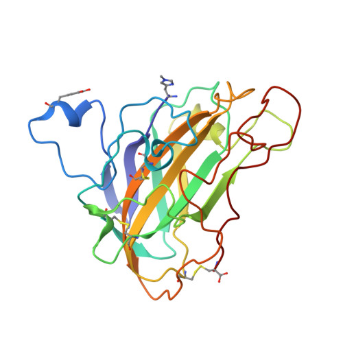
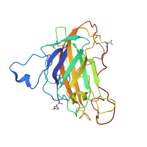
Structural basis for substrate targeting and catalysis by fungal polysaccharide monooxygenases.

Li, X.Beeson, W.T.Phillips, C.M.Marletta, M.A.Cate, J.H.

(2012) Structure 20: 1051-1061

  • DOI: https://doi.org/10.1016/j.str.2012.04.002
  • Primary Citation Related Structures: 
    4EIR, 4EIS

  • PubMed Abstract: 

    The use of cellulases remains a major cost in the production of renewable fuels and chemicals from lignocellulosic biomass. Fungi secrete copper-dependent polysaccharide monooxygenases (PMOs) that oxidatively cleave crystalline cellulose and improve the effectiveness of cellulases. However, the means by which PMOs recognize and cleave their substrates in the plant cell wall remain unclear. Here, we present structures of Neurospora crassa PMO-2 and PMO-3 at 1.10 and 1.37 Å resolution, respectively. In the structures, dioxygen species are found in the active sites, consistent with the proposed cleavage mechanism. Structural and sequence comparisons between PMOs also reveal that the enzyme substrate-binding surfaces contain highly varied aromatic amino acid and glycosylation positions. The structures reported here provide evidence for a wide range of PMO substrate recognition patterns in the plant cell wall, including binding modes that traverse multiple glucan chains.


  • Organizational Affiliation
    • California Institute for Quantitative Biosciences, University of California, Berkeley, CA 94720, USA.

Macromolecule Content 

  • Total Structure Weight: 49.55 kDa 
  • Atom Count: 4,246 
  • Modeled Residue Count: 450 
  • Deposited Residue Count: 450 
  • Unique protein chains: 2

Macromolecules

Find similar proteins by:|  3D Structure
Entity ID: 1
MoleculeChains  Sequence LengthOrganismDetailsImage
polysaccharide monooxygenase-3225Neurospora crassa OR74AMutation(s): 0 
EC: 1.14.99.56
UniProt
Find proteins for Q7SA19 (Neurospora crassa (strain ATCC 24698 / 74-OR23-1A / CBS 708.71 / DSM 1257 / FGSC 987))
Explore Q7SA19 
Go to UniProtKB:  Q7SA19
Entity Groups
Sequence Clusters30% Identity50% Identity70% Identity90% Identity95% Identity100% Identity
UniProt GroupQ7SA19
Glycosylation
Glycosylation Sites: 1
Sequence Annotations
Expand
Reference Sequence
Find similar proteins by:|  3D Structure
Entity ID: 2
MoleculeChains  Sequence LengthOrganismDetailsImage
polysaccharide monooxygenase-3225Neurospora crassa OR74AMutation(s): 0 
EC: 1.14.99.56
UniProt
Find proteins for Q7SA19 (Neurospora crassa (strain ATCC 24698 / 74-OR23-1A / CBS 708.71 / DSM 1257 / FGSC 987))
Explore Q7SA19 
Go to UniProtKB:  Q7SA19
Entity Groups
Sequence Clusters30% Identity50% Identity70% Identity90% Identity95% Identity100% Identity
UniProt GroupQ7SA19
Glycosylation
Glycosylation Sites: 1
Sequence Annotations
Expand
Reference Sequence

Oligosaccharides

Help  
Entity ID: 3
MoleculeChains Length2D Diagram GlycosylationD Interactions
2-acetamido-2-deoxy-beta-D-glucopyranose-(1-4)-2-acetamido-2-deoxy-beta-D-glucopyranose
C, D
2N-Glycosylation
Glycosylation Resources
GlyTouCan: G42666HT
GlyCosmos: G42666HT
GlyGen: G42666HT

Small Molecules

Experimental Data & Validation

Experimental Data

  • Method: X-RAY DIFFRACTION
  • Resolution: 1.37 Å
  • R-Value Free:  0.149 (Depositor), 0.151 (DCC) 
  • R-Value Work:  0.116 (Depositor), 0.121 (DCC) 
  • R-Value Observed: 0.118 (Depositor) 
Space Group: P 1 21 1
Unit Cell:
Length ( Å )Angle ( ˚ )
a = 35.74α = 90
b = 77.89β = 90.02
c = 82.05γ = 90
Software Package:
Software NamePurpose
REFMACrefinement
PDB_EXTRACTdata extraction
Blu-Icedata collection
MOSFLMdata reduction
SCALAdata scaling
PHASESphasing

Structure Validation

View Full Validation Report


Entry History 

Deposition Data

Revision History  (Full details and data files)

  • Version 1.0: 2012-05-23
    Type: Initial release
  • Version 1.1: 2012-06-27
    Changes: Database references
  • Version 2.0: 2020-07-29
    Type: Remediation
    Reason: Carbohydrate remediation
    Changes: Atomic model, Data collection, Database references, Derived calculations, Structure summary
  • Version 2.1: 2025-03-26
    Changes: Data collection, Database references, Structure summary