4DWR | pdb_00004dwr

RNA ligase RtcB/Mn2+ complex


Experimental Data Snapshot

  • Method: X-RAY DIFFRACTION
  • Resolution: 1.48 Å
  • R-Value Free: 
    0.178 (Depositor), 0.175 (DCC) 
  • R-Value Work: 
    0.142 (Depositor), 0.139 (DCC) 
  • R-Value Observed: 
    0.144 (Depositor) 

wwPDB Validation 3D Report Full Report

Validation slider image for 4DWR

This is version 2.1 of the entry. See complete history

Literature

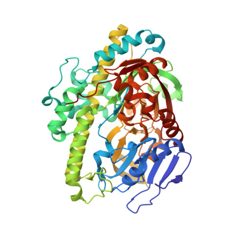
Structural and mechanistic insights into guanylylation of RNA-splicing ligase RtcB joining RNA between 3'-terminal phosphate and 5'-OH.

Englert, M.Xia, S.Okada, C.Nakamura, A.Tanavde, V.Yao, M.Eom, S.H.Konigsberg, W.H.Soll, D.Wang, J.

(2012) Proc Natl Acad Sci U S A 109: 15235-15240

  • DOI: https://doi.org/10.1073/pnas.1213795109
  • Primary Citation Related Structures: 
    4DWQ, 4DWR

  • PubMed Abstract: 

    The RtcB protein has recently been identified as a 3'-phosphate RNA ligase that directly joins an RNA strand ending with a 2',3'-cyclic phosphate to the 5'-hydroxyl group of another RNA strand in a GTP/Mn(2+)-dependent reaction. Here, we report two crystal structures of Pyrococcus horikoshii RNA-splicing ligase RtcB in complex with Mn(2+) alone (RtcB/ Mn(2+)) and together with a covalently bound GMP (RtcB-GMP/Mn(2+)). The RtcB/ Mn(2+) structure (at 1.6 Å resolution) shows two Mn(2+) ions at the active site, and an array of sulfate ions nearby that indicate the binding sites of the RNA phosphate backbone. The structure of the RtcB-GMP/Mn(2+) complex (at 2.3 Å resolution) reveals the detailed geometry of guanylylation of histidine 404. The critical roles of the key residues involved in the binding of the two Mn(2+) ions, the four sulfates, and GMP are validated in extensive mutagenesis and biochemical experiments, which also provide a thorough characterization for the three steps of the RtcB ligation pathway: (i) guanylylation of the enzyme, (ii) guanylyl-transfer to the RNA substrate, and (iii) overall ligation. These results demonstrate that the enzyme's substrate-induced GTP binding site and the putative reactive RNA ends are in the vicinity of the binuclear Mn(2+) active center, which provides detailed insight into how the enzyme-bound GMP is tansferred to the 3'-phosphate of the RNA substrate for activation and subsequent nucleophilic attack by the 5'-hydroxyl of the second RNA substrate, resulting in the ligated product and release of GMP.


  • Organizational Affiliation
    • Department of Molecular Biophysics and Biochemistry, Yale University, New Haven, CT 06520, USA.

Macromolecule Content 

  • Total Structure Weight: 165.9 kDa 
  • Atom Count: 13,603 
  • Modeled Residue Count: 1,450 
  • Deposited Residue Count: 1,461 
  • Unique protein chains: 1

Macromolecules

Find similar proteins by:|  3D Structure
Entity ID: 1
MoleculeChains  Sequence LengthOrganismDetailsImage
tRNA-splicing ligase RtcBA [auth B],
B [auth A],
C
487Pyrococcus horikoshiiMutation(s): 0 
EC: 6.5.1 (PDB Primary Data), 6.5.1.8 (UniProt)
UniProt
Find proteins for O59245 (Pyrococcus horikoshii (strain ATCC 700860 / DSM 12428 / JCM 9974 / NBRC 100139 / OT-3))
Explore O59245 
Go to UniProtKB:  O59245
Entity Groups
Sequence Clusters30% Identity50% Identity70% Identity90% Identity95% Identity100% Identity
UniProt GroupO59245
Sequence Annotations
Expand
Reference Sequence

Oligosaccharides

Help  
Entity ID: 2
MoleculeChains Length2D Diagram GlycosylationD Interactions
beta-D-fructofuranose-(2-1)-alpha-D-glucopyranose
D, E, F
2N/A
Glycosylation Resources
GlyTouCan: G05551OP
GlyCosmos: G05551OP

Small Molecules

Ligands 2 Unique
IDChains Name / Formula / InChI Key2D Diagram3D Interactions
SO4

Query on SO4


Download:Ideal Coordinates CCD File
AA [auth C]
BA [auth C]
CA [auth C]
DA [auth C]
EA [auth C]
AA [auth C],
BA [auth C],
CA [auth C],
DA [auth C],
EA [auth C],
FA [auth C],
GA [auth C],
I [auth B],
J [auth B],
K [auth B],
L [auth B],
M [auth B],
N [auth B],
O [auth B],
P [auth B],
S [auth A],
T [auth A],
U [auth A],
V [auth A],
W [auth A],
X [auth A]
SULFATE ION
O4 S
QAOWNCQODCNURD-UHFFFAOYSA-L
MN

Query on MN


Download:Ideal Coordinates CCD File
G [auth B]
H [auth B]
Q [auth A]
R [auth A]
Y [auth C]
G [auth B],
H [auth B],
Q [auth A],
R [auth A],
Y [auth C],
Z [auth C]
MANGANESE (II) ION
Mn
WAEMQWOKJMHJLA-UHFFFAOYSA-N

Biologically Interesting Molecules (External Reference) 

1 Unique

Experimental Data & Validation

Experimental Data

  • Method: X-RAY DIFFRACTION
  • Resolution: 1.48 Å
  • R-Value Free:  0.178 (Depositor), 0.175 (DCC) 
  • R-Value Work:  0.142 (Depositor), 0.139 (DCC) 
  • R-Value Observed: 0.144 (Depositor) 
Space Group: P 1 21 1
Unit Cell:
Length ( Å )Angle ( ˚ )
a = 114.081α = 90
b = 84.899β = 115.65
c = 124.2γ = 90
Software Package:
Software NamePurpose
HKL-2000data collection
AMoREphasing
REFMACrefinement
HKL-2000data reduction
HKL-2000data scaling

Structure Validation

View Full Validation Report


Entry History 

Deposition Data

Revision History  (Full details and data files)

  • Version 1.0: 2012-09-05
    Type: Initial release
  • Version 1.1: 2012-09-26
    Changes: Database references
  • Version 1.2: 2012-12-19
    Changes: Database references
  • Version 1.3: 2017-08-02
    Changes: Refinement description, Source and taxonomy
  • Version 2.0: 2020-07-29
    Type: Remediation
    Reason: Carbohydrate remediation
    Changes: Atomic model, Data collection, Database references, Derived calculations, Non-polymer description, Structure summary
  • Version 2.1: 2024-02-28
    Changes: Data collection, Database references, Structure summary