4DJ4 | pdb_00004dj4

X-ray structure of mutant N211D of bifunctional nuclease TBN1 from Solanum lycopersicum (Tomato)


Experimental Data Snapshot

  • Method: X-RAY DIFFRACTION
  • Resolution: 2.35 Å
  • R-Value Free: 
    0.207 (Depositor), 0.187 (DCC) 
  • R-Value Work: 
    0.183 (Depositor), 0.187 (DCC) 
  • R-Value Observed: 
    0.186 (Depositor) 

Starting Model: experimental
View more details

wwPDB Validation 3D Report Full Report

Validation slider image for 4DJ4

This is version 2.2 of the entry. See complete history

Literature

Plant multifunctional nuclease TBN1 with unexpected phospholipase activity: structural study and reaction-mechanism analysis.

Koval, T.Lipovova, P.Podzimek, T.Matousek, J.Duskova, J.Skalova, T.Stepankova, A.Hasek, J.Dohnalek, J.

(2013) Acta Crystallogr D Biol Crystallogr 69: 213-226

  • DOI: https://doi.org/10.1107/S0907444912043697
  • Primary Citation Related Structures: 
    3SNG, 4DJ4

  • PubMed Abstract: 

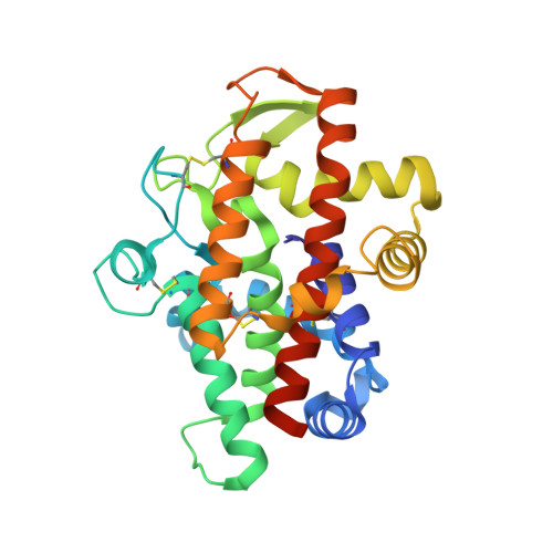
    Type I plant nucleases play an important role in apoptotic processes and cell senescence. Recently, they have also been indicated to be potent anticancer agents in in vivo studies. The first structure of tomato nuclease I (TBN1) has been determined, its oligomerization and activity profiles have been analyzed and its unexpected activity towards phospholipids has been discovered, and conclusions are drawn regarding its catalytic mechanism. The structure-solution process required X-ray diffraction data from two crystal forms. The first form was used for phase determination; the second form was used for model building and refinement. TBN1 is mainly α-helical and is stabilized by four disulfide bridges. Three observed oligosaccharides are crucial for its stability and solubility. The active site is localized at the bottom of the positively charged groove and contains a zinc cluster that is essential for enzymatic activity. An equilibrium between monomers, dimers and higher oligomers of TBN1 was observed in solution. Principles of the reaction mechanism of the phosphodiesterase activity are suggested, with central roles for the zinc cluster, the nucleobase-binding pocket (Phe-site) and Asp70, Arg73 and Asn167. Based on the distribution of surface residues, possible binding sites for dsDNA and other nucleic acids with secondary structure were identified. The phospholipase activity of TBN1, which is reported for the first time for a nuclease, significantly broadens the substrate promiscuity of the enzyme, and the resulting release of diacylglycerol, which is an important second messenger, can be related to the role of TBN1 in apoptosis.


  • Organizational Affiliation
    • Institute of Macromolecular Chemistry, AS CR, v.v.i., Heyrovskeho nam. 2, 162 06 Praha 6, Czech Republic. koval.tomas@gmail.com

Macromolecule Content 

  • Total Structure Weight: 66.78 kDa 
  • Atom Count: 4,593 
  • Modeled Residue Count: 517 
  • Deposited Residue Count: 554 
  • Unique protein chains: 1

Macromolecules

Find similar proteins by:|  3D Structure
Entity ID: 1
MoleculeChains  Sequence LengthOrganismDetailsImage
Nuclease
A, B
277Solanum lycopersicumMutation(s): 1 
Gene Names: tbn1
EC: 3.1.30.1
UniProt
Find proteins for Q0KFV0 (Solanum lycopersicum)
Explore Q0KFV0 
Go to UniProtKB:  Q0KFV0
Entity Groups
Sequence Clusters30% Identity50% Identity70% Identity90% Identity95% Identity100% Identity
UniProt GroupQ0KFV0
Glycosylation
Glycosylation Sites: 2
Sequence Annotations
Expand
Reference Sequence

Oligosaccharides

Help  
Entity ID: 2
MoleculeChains Length2D Diagram GlycosylationD Interactions
2-acetamido-2-deoxy-beta-D-glucopyranose-(1-4)-2-acetamido-2-deoxy-beta-D-glucopyranose
C
2N-Glycosylation
Glycosylation Resources
GlyTouCan: G42666HT
GlyCosmos: G42666HT
GlyGen: G42666HT
Entity ID: 3
MoleculeChains Length2D Diagram GlycosylationD Interactions
alpha-D-mannopyranose-(1-3)-[alpha-D-mannopyranose-(1-6)]beta-D-mannopyranose-(1-4)-2-acetamido-2-deoxy-beta-D-glucopyranose-(1-4)-2-acetamido-2-deoxy-beta-D-glucopyranose
D, F
5N-Glycosylation
Glycosylation Resources
GlyTouCan: G22768VO
GlyCosmos: G22768VO
GlyGen: G22768VO
Entity ID: 4
MoleculeChains Length2D Diagram GlycosylationD Interactions
alpha-D-mannopyranose-(1-3)-beta-D-mannopyranose-(1-4)-2-acetamido-2-deoxy-beta-D-glucopyranose-(1-4)-2-acetamido-2-deoxy-beta-D-glucopyranose
E
4N-Glycosylation
Glycosylation Resources
GlyTouCan: G81315DD
GlyCosmos: G81315DD
GlyGen: G81315DD

Small Molecules

Ligands 3 Unique
IDChains Name / Formula / InChI Key2D Diagram3D Interactions
ZN

Query on ZN


Download:Ideal Coordinates CCD File
G [auth A]
H [auth A]
I [auth A]
K [auth B]
L [auth B]
G [auth A],
H [auth A],
I [auth A],
K [auth B],
L [auth B],
M [auth B]
ZINC ION
Zn
PTFCDOFLOPIGGS-UHFFFAOYSA-N
CL

Query on CL


Download:Ideal Coordinates CCD File
J [auth A],
N [auth B]
CHLORIDE ION
Cl
VEXZGXHMUGYJMC-UHFFFAOYSA-M
NA

Query on NA


Download:Ideal Coordinates CCD File
O [auth B]SODIUM ION
Na
FKNQFGJONOIPTF-UHFFFAOYSA-N

Experimental Data & Validation

Experimental Data

  • Method: X-RAY DIFFRACTION
  • Resolution: 2.35 Å
  • R-Value Free:  0.207 (Depositor), 0.187 (DCC) 
  • R-Value Work:  0.183 (Depositor), 0.187 (DCC) 
  • R-Value Observed: 0.186 (Depositor) 
Space Group: H 3
Unit Cell:
Length ( Å )Angle ( ˚ )
a = 113.322α = 90
b = 113.322β = 90
c = 138.322γ = 120
Software Package:
Software NamePurpose
MxCuBEdata collection
MOLREPphasing
REFMACrefinement
HKL-2000data reduction
HKL-2000data scaling

Structure Validation

View Full Validation Report


Entry History 

Deposition Data

Revision History  (Full details and data files)

  • Version 1.0: 2012-11-07
    Type: Initial release
  • Version 1.1: 2013-02-20
    Changes: Database references
  • Version 1.2: 2018-01-31
    Changes: Database references
  • Version 2.0: 2020-07-29
    Type: Remediation
    Reason: Carbohydrate remediation
    Changes: Atomic model, Data collection, Database references, Derived calculations, Structure summary
  • Version 2.1: 2023-09-13
    Changes: Data collection, Database references, Refinement description, Structure summary
  • Version 2.2: 2024-11-27
    Changes: Structure summary