4C64 | pdb_00004c64

ULTRA HIGH RESOLUTION DICKERSON-DREW DODECAMER B-DNA


Experimental Data Snapshot

  • Method: X-RAY DIFFRACTION
  • Resolution: 1.32 Å
  • R-Value Free: 
    0.182 (Depositor), 0.185 (DCC) 
  • R-Value Work: 
    0.139 (Depositor), 0.145 (DCC) 
  • R-Value Observed: 
    0.143 (Depositor) 

Starting Model: experimental
View more details

wwPDB Validation 3D Report Full Report

Validation slider image for 4C64

This is version 1.4 of the entry. See complete history

Literature

Structural insights into how 5-hydroxymethylation influences transcription factor binding.

Lercher, L.McDonough, M.A.El-Sagheer, A.H.Thalhammer, A.Kriaucionis, S.Brown, T.Schofield, C.J.

(2014) Chem Commun (Camb) 50: 1794-1796

  • DOI: https://doi.org/10.1039/c3cc48151d
  • Primary Citation Related Structures: 
    4C5X, 4C63, 4C64

  • PubMed Abstract: 

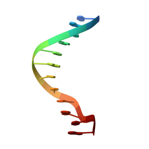
    Transcription factor binding and high resolution crystallographic studies (1.3 Å) of Dickerson-Drew duplexes with cytosine, methylcytosine and hydroxymethylcytosine bases provide evidence that C-5 cytosine modifications could regulate transcription by context dependent effects on DNA transcription factor interactions.


  • Organizational Affiliation
    • Department of Chemistry and the Oxford Centre for Integrative Systems Biology, Chemistry Research Laboratory, Oxford, UK. Christopher.schofield@chem.ox.ac.uk.

Macromolecule Content 

  • Total Structure Weight: 7.35 kDa 
  • Atom Count: 618 
  • Modeled Residue Count: 24 
  • Deposited Residue Count: 24 
  • Unique nucleic acid chains: 1

Macromolecules

Find similar nucleic acids by:  Sequence
Entity ID: 1
MoleculeChains LengthOrganismImage
5'-D(*CP*GP*CP*GP*AP*AP*TP*TP*CP*GP*CP*GP)-3'
A, B
12synthetic construct
Sequence Annotations
Expand
Reference Sequence

Small Molecules

Ligands 1 Unique
IDChains Name / Formula / InChI Key2D Diagram3D Interactions
MG

Query on MG


Download:Ideal Coordinates CCD File
C [auth A]MAGNESIUM ION
Mg
JLVVSXFLKOJNIY-UHFFFAOYSA-N

Experimental Data & Validation

Experimental Data

  • Method: X-RAY DIFFRACTION
  • Resolution: 1.32 Å
  • R-Value Free:  0.182 (Depositor), 0.185 (DCC) 
  • R-Value Work:  0.139 (Depositor), 0.145 (DCC) 
  • R-Value Observed: 0.143 (Depositor) 
Space Group: P 21 21 21
Unit Cell:
Length ( Å )Angle ( ˚ )
a = 25.149α = 90
b = 39.84β = 90
c = 65.598γ = 90
Software Package:
Software NamePurpose
PHENIXrefinement
XDSdata reduction
SCALAdata scaling
PHASERphasing

Structure Validation

View Full Validation Report


Entry History 

Deposition Data

Revision History  (Full details and data files)

  • Version 1.0: 2013-10-02
    Type: Initial release
  • Version 1.1: 2013-12-11
    Changes: Database references
  • Version 1.2: 2014-02-12
    Changes: Database references
  • Version 1.3: 2018-02-21
    Changes: Database references
  • Version 1.4: 2023-12-20
    Changes: Data collection, Database references, Derived calculations, Other, Refinement description