4BX2

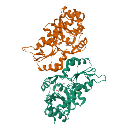
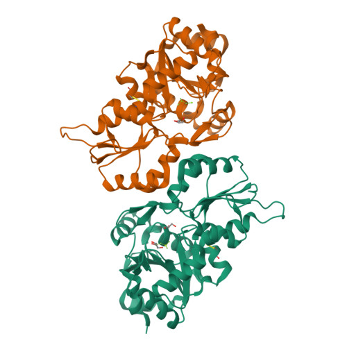
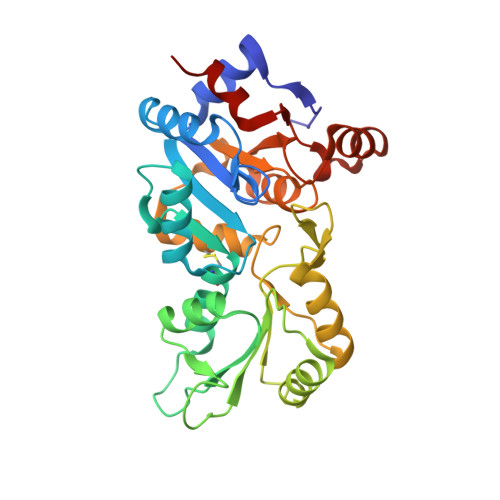
Crystal Structure of murine Chronophin (Pyridoxal Phosphate Phosphatase) in complex with Beryllium trifluoride


Experimental Data Snapshot

  • Method: X-RAY DIFFRACTION
  • Resolution: 2.19 Å
  • R-Value Free: 0.212 
  • R-Value Work: 0.168 
  • R-Value Observed: 0.170 

Starting Model: experimental
View more details

wwPDB Validation   3D Report Full Report


This is version 2.1 of the entry. See complete history


Literature

Chronophin Dimerization is Required for Proper Positioning of its Substrate Specificity Loop.

Kestler, C.Knobloch, G.Tessmer, I.Jeanclos, E.Schindelin, H.Gohla, A.

(2014) J Biol Chem 289: 3094

  • DOI: https://doi.org/10.1074/jbc.M113.536482
  • Primary Citation of Related Structures:  
    4BX0, 4BX2, 4BX3

  • PubMed Abstract: 

    Mammalian phosphatases of the haloacid dehalogenase (HAD) superfamily have emerged as important regulators of physiology and disease. Many of these enzymes are stable homodimers; however, the role of their dimerization is largely unknown. Here, we explore the function of the obligatory homodimerization of chronophin, a mammalian HAD phosphatase known to dephosphorylate pyridoxal 5'-phosphate (PLP) and serine/threonine-phosphorylated proteins. The exchange of two residues in the murine chronophin homodimerization interface (chronophin(A194K,A195K)) yields a constitutive monomer both in vitro and in cells. The catalytic activity of monomeric chronophin toward PLP is strongly impaired. X-ray crystallographic studies of chronophin(A194K,A195K) revealed that dimer formation is essential for an intermolecular arginine-arginine-tryptophan stacking interaction that positions a critical histidine residue in the substrate specificity loop of chronophin for PLP coordination. Analysis of all available crystal structures of HAD hydrolases that are grouped together with chronophin in the C2a-type structural subfamily uncovered a highly conserved mode of dimerization that results in intermolecular contacts involving the substrate specificity loop. Our results explain how the dimerization of HAD hydrolases contributes to their catalytic efficiency and substrate specificity.


  • Organizational Affiliation

    From the Institute for Pharmacology and Toxicology, University of Würzburg, 97078 Würzburg, Germany and.


Macromolecules
Find similar proteins by:  (by identity cutoff)  |  3D Structure
Entity ID: 1
MoleculeChains Sequence LengthOrganismDetailsImage
PYRIDOXAL PHOSPHATE PHOSPHATASE
A, B
293Mus musculusMutation(s): 0 
EC: 3.1.3.74 (PDB Primary Data), 3.1.3.3 (PDB Primary Data), 3.1.3.16 (UniProt)
UniProt & NIH Common Fund Data Resources
Find proteins for P60487 (Mus musculus)
Explore P60487 
Go to UniProtKB:  P60487
IMPC:  MGI:1919282
Entity Groups  
Sequence Clusters30% Identity50% Identity70% Identity90% Identity95% Identity100% Identity
UniProt GroupP60487
Sequence Annotations
Expand
  • Reference Sequence
Small Molecules
Ligands 3 Unique
IDChains Name / Formula / InChI Key2D Diagram3D Interactions
GOL
Query on GOL

Download Ideal Coordinates CCD File 
E [auth A],
F [auth A],
I [auth B]
GLYCEROL
C3 H8 O3
PEDCQBHIVMGVHV-UHFFFAOYSA-N
BEF
Query on BEF

Download Ideal Coordinates CCD File 
D [auth A],
H [auth B]
BERYLLIUM TRIFLUORIDE ION
Be F3
OGIAHMCCNXDTIE-UHFFFAOYSA-K
MG
Query on MG

Download Ideal Coordinates CCD File 
C [auth A],
G [auth B]
MAGNESIUM ION
Mg
JLVVSXFLKOJNIY-UHFFFAOYSA-N
Experimental Data & Validation

Experimental Data

  • Method: X-RAY DIFFRACTION
  • Resolution: 2.19 Å
  • R-Value Free: 0.212 
  • R-Value Work: 0.168 
  • R-Value Observed: 0.170 
  • Space Group: I 2 3
Unit Cell:
Length ( Å )Angle ( ˚ )
a = 166.83α = 90
b = 166.83β = 90
c = 166.83γ = 90
Software Package:
Software NamePurpose
PHENIXrefinement
iMOSFLMdata reduction
SCALAdata scaling
PHASERphasing

Structure Validation

View Full Validation Report


Entry History 

Deposition Data

Revision History  (Full details and data files)

  • Version 1.0: 2013-12-25
    Type: Initial release
  • Version 1.1: 2014-02-12
    Changes: Database references
  • Version 2.0: 2023-12-20
    Changes: Atomic model, Data collection, Database references, Derived calculations, Other, Refinement description
  • Version 2.1: 2024-10-23
    Changes: Structure summary