4B20 | pdb_00004b20

Structural basis of DNA loop recognition by Endonuclease V


Experimental Data Snapshot

  • Method: X-RAY DIFFRACTION
  • Resolution: 2.75 Å
  • R-Value Free: 
    0.291 (Depositor), 0.279 (DCC) 
  • R-Value Work: 
    0.227 (Depositor), 0.219 (DCC) 
  • R-Value Observed: 
    0.227 (Depositor) 

wwPDB Validation 3D Report Full Report

Validation slider image for 4B20

This is version 1.2 of the entry. See complete history

Literature

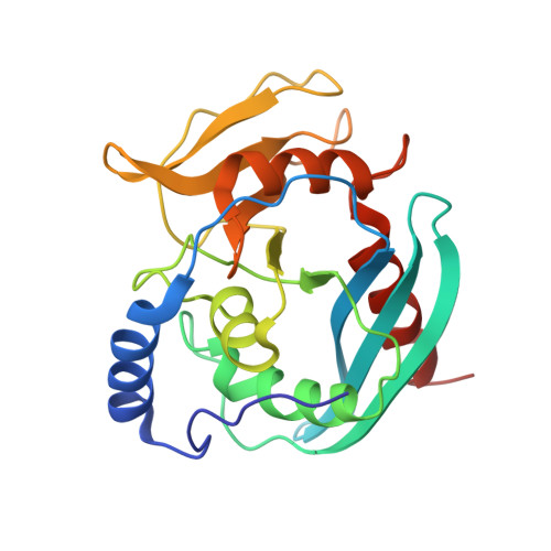
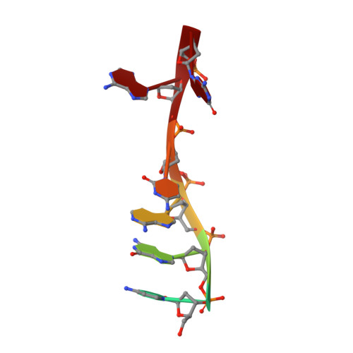
Structural Basis of DNA Loop Recognition by Endonuclease V.

Rosnes, I.Rowe, A.D.Vik, E.S.Forstrom, R.J.Alseth, I.Bjoras, M.Dalhus, B.

(2013) Structure 21: 257

  • DOI: https://doi.org/10.1016/j.str.2012.12.007
  • Primary Citation Related Structures: 
    4B20

  • PubMed Abstract: 

    The DNA repair enzyme endonuclease V (EndoV) recognizes and cleaves DNA at deaminated adenine lesions (hypoxanthine). In addition, EndoV cleaves DNA containing various helical distortions such as loops, hairpins, and flaps. To understand the molecular basis of EndoV's ability to recognize and incise DNA structures with helical distortions, we solved the crystal structure of Thermotoga maritima EndoV in complex with DNA containing a one-nucleotide loop. The structure shows that a strand-separating wedge is crucial for DNA loop recognition, with DNA strands separated precisely at the helical distortion. The additional nucleotide forming the loop rests on the surface of the wedge, while the normal adenine opposite the loop is flipped into a base recognition pocket. Our data show a different principle for DNA loop recognition and cleavage by EndoV, in which a coordinated action of a DNA-intercalating wedge and a base pocket accommodating a flipped normal base facilitate strand incision.


  • Organizational Affiliation
    • Department of Microbiology, Oslo University Hospital, Rikshospitalet, P.O. Box 4950, Nydalen, N-0424 Oslo, Norway.

Macromolecule Content 

  • Total Structure Weight: 61.64 kDa 
  • Atom Count: 4,290 
  • Modeled Residue Count: 479 
  • Deposited Residue Count: 484 
  • Unique protein chains: 1
  • Unique nucleic acid chains: 2

Macromolecules


Find similar proteins by:|  3D Structure
Entity ID: 1
MoleculeChains  Sequence LengthOrganismDetailsImage
Endonuclease V
A, B
225Thermotoga maritimaMutation(s): 0 
Gene Names: nfiTM_1865
EC: 3.1.21.7
UniProt
Find proteins for Q9X2H9 (Thermotoga maritima (strain ATCC 43589 / DSM 3109 / JCM 10099 / NBRC 100826 / MSB8))
Explore Q9X2H9 
Go to UniProtKB:  Q9X2H9
Entity Groups
Sequence Clusters30% Identity50% Identity70% Identity90% Identity95% Identity100% Identity
UniProt GroupQ9X2H9
Sequence Annotations
Expand
Reference Sequence
Find similar nucleic acids by:  Sequence
Entity ID: 2
MoleculeChains LengthOrganismImage
5'-D(*GP*CP*GP*AP*CP*AP*GP)-3'C,
E [auth F]
7N/A
Sequence Annotations
Expand
Reference Sequence
Find similar nucleic acids by:  Sequence
Entity ID: 3
MoleculeChains LengthOrganismImage
5'-D(*AP*TP*CP*TP*TP*GP*TP*CP*GP*CP)-3'D,
F [auth G]
10N/A
Sequence Annotations
Expand
Reference Sequence

Experimental Data & Validation

Experimental Data

  • Method: X-RAY DIFFRACTION
  • Resolution: 2.75 Å
  • R-Value Free:  0.291 (Depositor), 0.279 (DCC) 
  • R-Value Work:  0.227 (Depositor), 0.219 (DCC) 
  • R-Value Observed: 0.227 (Depositor) 
Space Group: I 2 2 2
Unit Cell:
Length ( Å )Angle ( ˚ )
a = 55.231α = 90
b = 135.37β = 90
c = 194.993γ = 90
Software Package:
Software NamePurpose
CNSrefinement
MOSFLMdata reduction
SCALAdata scaling
CNSphasing

Structure Validation

View Full Validation Report


Entry History 

Deposition Data

Revision History  (Full details and data files)

  • Version 1.0: 2013-04-17
    Type: Initial release
  • Version 1.1: 2024-05-08
    Changes: Data collection, Database references, Derived calculations, Other
  • Version 1.2: 2025-12-10
    Changes: Database references, Derived calculations, Source and taxonomy, Structure summary