3D34 | pdb_00003d34

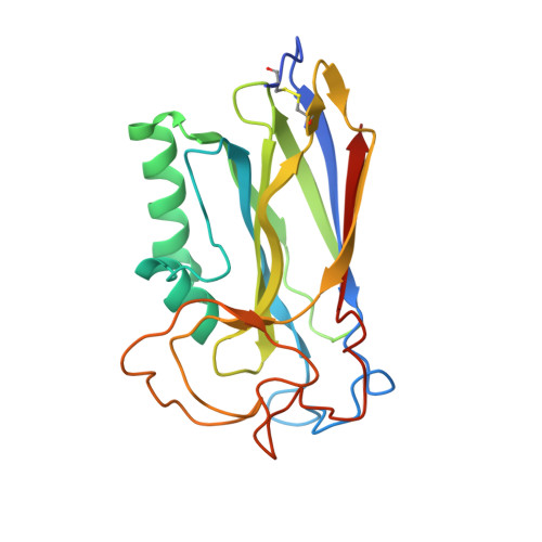
Structure of the F-spondin domain of mindin


Experimental Data Snapshot

  • Method: X-RAY DIFFRACTION
  • Resolution: 1.80 Å
  • R-Value Free: 
    0.215 (Depositor), 0.211 (DCC) 
  • R-Value Work: 
    0.189 (Depositor), 0.184 (DCC) 

wwPDB Validation 3D Report Full Report

Validation slider image for 3D34

This is version 1.3 of the entry. See complete history. 

Literature

Structure of the F-spondin domain of mindin, an integrin ligand and pattern recognition molecule.

Li, Y., Cao, C., Jia, W., Yu, L., Mo, M., Wang, Q., Huang, Y., Lim, J.M., Ishihara, M., Wells, L., Azadi, P., Robinson, H., He, Y.W., Zhang, L., Mariuzza, R.A.

(2009) EMBO J 28: 286-297

  • DOI: https://doi.org/10.1038/emboj.2008.288
  • Primary Citation Related Structures: 
    3D34

  • PubMed Abstract: 

    Mindin (spondin-2) is an extracellular matrix protein of unknown structure that is required for efficient T-cell priming by dendritic cells. Additionally, mindin functions as a pattern recognition molecule for initiating innate immune responses. These dual functions are mediated by interactions with integrins and microbial pathogens, respectively. Mindin comprises an N-terminal F-spondin (FS) domain and C-terminal thrombospondin type 1 repeat (TSR). We determined the structure of the FS domain at 1.8-A resolution. The structure revealed an eight-stranded antiparallel beta-sandwich motif resembling that of membrane-targeting C2 domains, including a bound calcium ion. We demonstrated that the FS domain mediates integrin binding and identified the binding site by mutagenesis. The mindin FS domain therefore represents a new integrin ligand. We further showed that mindin recognizes lipopolysaccharide (LPS) through its TSR domain, and obtained evidence that C-mannosylation of the TSR influences LPS binding. Through these dual interactions, the FS and TSR domains of mindin promote activation of both adaptive and innate immune responses.


  • Organizational Affiliation: 
    • WM Keck Laboratory for Structural Biology, Center for Advanced Research in Biotechnology, University of Maryland Biotechnology Institute, Rockville, MD 20850, USA.

Macromolecule Content 

  • Total Structure Weight: 49.77 kDa 
  • Atom Count: 3,606 
  • Modeled Residue Count: 426 
  • Deposited Residue Count: 446 
  • Unique protein chains: 1

Macromolecules

Find similar proteins by:|  3D Structure
Entity ID: 1
MoleculeChains  Sequence LengthOrganismDetailsImage
Spondin-2
A, B
223Homo sapiensMutation(s): 0 
Gene Names: SPON2, DIL1
Membrane Entity: Yes 
UniProt & NIH Common Fund Data Resources
Find proteins for Q9BUD6 (Homo sapiens)
Explore Q9BUD6 
Go to UniProtKB:  Q9BUD6
PHAROS:  Q9BUD6
GTEx:  ENSG00000159674 
Entity Groups
Sequence Clusters30% Identity50% Identity70% Identity90% Identity95% Identity100% Identity
UniProt GroupQ9BUD6
Sequence Annotations
Expand
Reference Sequence

Small Molecules

Ligands 2 Unique
IDChains Name / Formula / InChI Key2D Diagram3D Interactions
NI

Query on NI


Download:Ideal Coordinates CCD File
C [auth A]
D [auth A]
E [auth A]
F [auth A]
H [auth B]
C [auth A],
D [auth A],
E [auth A],
F [auth A],
H [auth B],
I [auth B]
NICKEL (II) ION
Ni
VEQPNABPJHWNSG-UHFFFAOYSA-N
CA

Query on CA


Download:Ideal Coordinates CCD File
G [auth A],
J [auth B]
CALCIUM ION
Ca
BHPQYMZQTOCNFJ-UHFFFAOYSA-N

Experimental Data & Validation

Experimental Data

  • Method: X-RAY DIFFRACTION
  • Resolution: 1.80 Å
  • R-Value Free:  0.215 (Depositor), 0.211 (DCC) 
  • R-Value Work:  0.189 (Depositor), 0.184 (DCC) 
Space Group: P 21 21 21
Unit Cell:
Length ( Å )Angle ( ˚ )
a = 50.6α = 90
b = 68.027β = 90
c = 110.13γ = 90
Software Package:
Software NamePurpose
d*TREKdata scaling
CNSrefinement
PDB_EXTRACTdata extraction
DENZOdata reduction
SCALEPACKdata scaling

Structure Validation

View Full Validation Report


Entry History 

Deposition Data

Revision History  (Full details and data files)

  • Version 1.0: 2009-02-17
    Type: Initial release
  • Version 1.1: 2011-07-13
    Changes: Version format compliance
  • Version 1.2: 2017-10-25
    Changes: Advisory, Refinement description
  • Version 1.3: 2024-10-30
    Changes: Advisory, Data collection, Database references, Derived calculations, Structure summary