3ZD7 | pdb_00003zd7

Snapshot 3 of RIG-I scanning on RNA duplex


Experimental Data Snapshot

  • Method: X-RAY DIFFRACTION
  • Resolution: 2.50 Å
  • R-Value Free: 
    0.288 (Depositor), 0.280 (DCC) 
  • R-Value Work: 
    0.229 (Depositor), 0.222 (DCC) 
  • R-Value Observed: 
    0.232 (Depositor) 

Starting Model: experimental
View more details

wwPDB Validation 3D Report Full Report

Validation slider image for 3ZD7

Ligand Structure Quality Assessment 


This is version 1.3 of the entry. See complete history

Literature

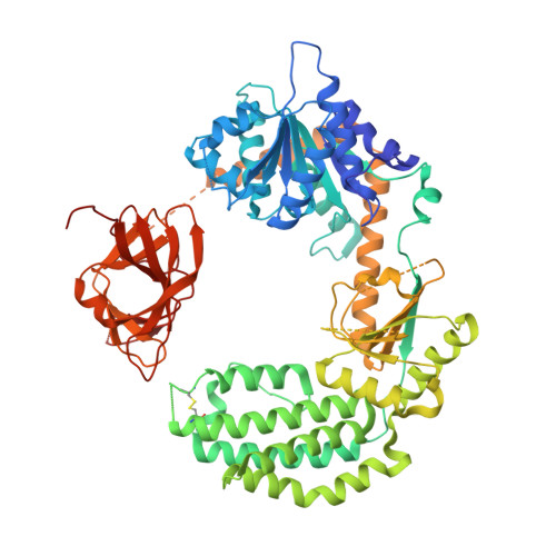
Defining the Functional Determinants for RNA Surveillance by Rig-I.

Kohlway, A.Luo, D.Rawling, D.C.Ding, S.C.Pyle, A.M.

(2013) EMBO Rep 14: 772

  • DOI: https://doi.org/10.1038/embor.2013.108
  • Primary Citation Related Structures: 
    3ZD6, 3ZD7

  • PubMed Abstract: 

    Retinoic acid-inducible gene-I (RIG-I) is an intracellular RNA sensor that activates the innate immune machinery in response to infection by RNA viruses. Here, we report the crystal structure of distinct conformations of a RIG-I:dsRNA complex, which shows that HEL2i-mediated scanning allows RIG-I to sense the length of RNA targets. To understand the implications of HEL2i scanning for catalytic activity and signalling by RIG-I, we examined its ATPase activity when stimulated by duplex RNAs of varying lengths and 5' composition. We identified a minimal RNA duplex that binds one RIG-I molecule, stimulates robust ATPase activity, and elicits a RIG-I-mediated interferon response in cells. Our results reveal that the minimal functional unit of the RIG-I:RNA complex is a monomer that binds at the terminus of a duplex RNA substrate. This behaviour is markedly different from the RIG-I paralog melanoma differentiation-associated gene 5 (MDA5), which forms cooperative filaments.


  • Organizational Affiliation
    • Department of Molecular Biophysics and Biochemistry.

Macromolecule Content 

  • Total Structure Weight: 86.64 kDa 
  • Atom Count: 5,369 
  • Modeled Residue Count: 643 
  • Deposited Residue Count: 716 
  • Unique protein chains: 1
  • Unique nucleic acid chains: 1

Macromolecules


Find similar proteins by:|  3D Structure
Entity ID: 1
MoleculeChains  Sequence LengthOrganismDetailsImage
PROBABLE ATP-DEPENDENT RNA HELICASE DDX58696Homo sapiensMutation(s): 0 
EC: 3.6.4.13
UniProt & NIH Common Fund Data Resources
Find proteins for O95786 (Homo sapiens)
Explore O95786 
Go to UniProtKB:  O95786
PHAROS:  O95786
GTEx:  ENSG00000107201 
Entity Groups
Sequence Clusters30% Identity50% Identity70% Identity90% Identity95% Identity100% Identity
UniProt GroupO95786
Sequence Annotations
Expand
Reference Sequence
Find similar nucleic acids by:  Sequence
Entity ID: 2
MoleculeChains LengthOrganismImage
RNA DUPLEXB [auth C],
C [auth D]
10synthetic construct
Sequence Annotations
Expand
Reference Sequence

Experimental Data & Validation

Experimental Data

  • Method: X-RAY DIFFRACTION
  • Resolution: 2.50 Å
  • R-Value Free:  0.288 (Depositor), 0.280 (DCC) 
  • R-Value Work:  0.229 (Depositor), 0.222 (DCC) 
  • R-Value Observed: 0.232 (Depositor) 
Space Group: P 21 21 21
Unit Cell:
Length ( Å )Angle ( ˚ )
a = 48.252α = 90
b = 75.996β = 90
c = 207.279γ = 90
Software Package:
Software NamePurpose
REFMACrefinement
PHASERphasing

Structure Validation

View Full Validation Report


Ligand Structure Quality Assessment 


Entry History 

Deposition Data

Revision History  (Full details and data files)

  • Version 1.0: 2013-08-07
    Type: Initial release
  • Version 1.1: 2013-09-11
    Changes: Database references
  • Version 1.2: 2023-12-20
    Changes: Data collection, Database references, Derived calculations, Other, Refinement description
  • Version 1.3: 2024-11-20
    Changes: Structure summary