3WWJ

Crystal structure of an engineered sitagliptin-producing transaminase, ATA-117-Rd11


Experimental Data Snapshot

  • Method: X-RAY DIFFRACTION
  • Resolution: 2.20 Å
  • R-Value Free: 0.213 
  • R-Value Work: 0.168 
  • R-Value Observed: 0.170 

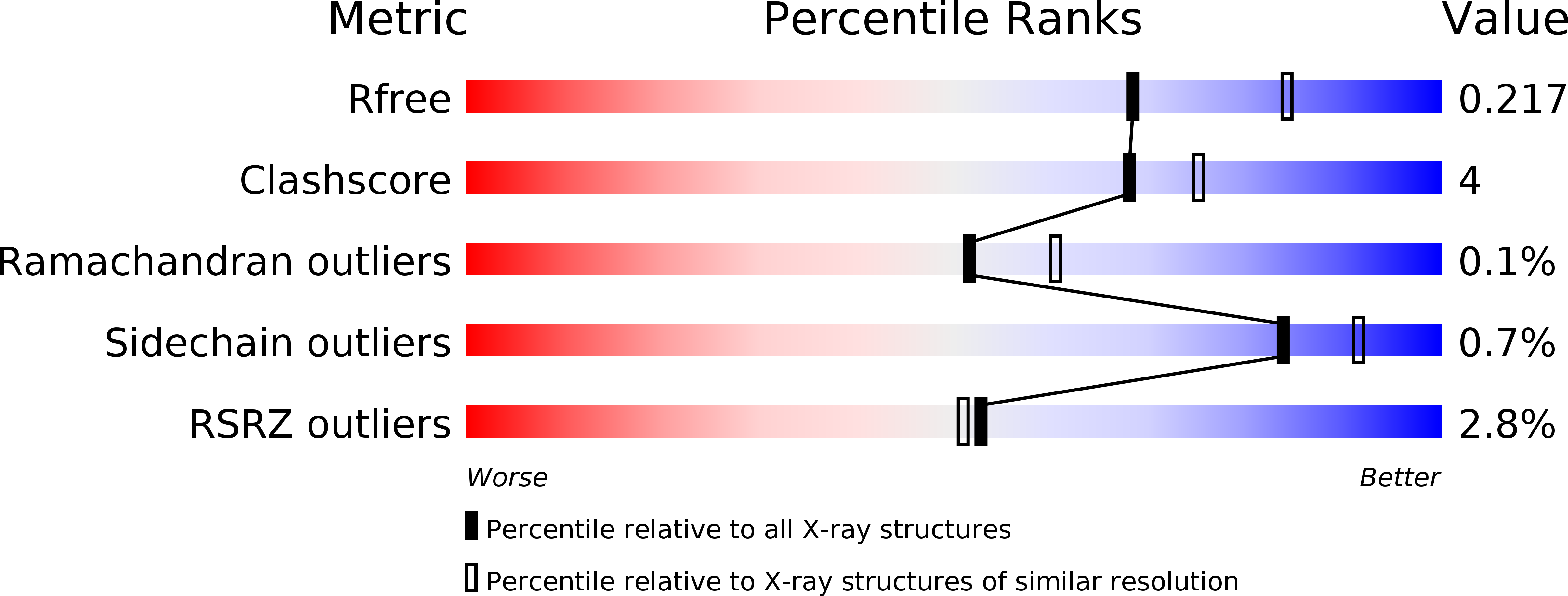
wwPDB Validation   3D Report Full Report


Ligand Structure Quality Assessment 


This is version 1.1 of the entry. See complete history


Literature

A new target region for changing the substrate specificity of amine transaminases.

Guan, L.J.Ohtsuka, J.Okai, M.Miyakawa, T.Mase, T.Zhi, Y.Hou, F.Ito, N.Iwasaki, A.Yasohara, Y.Tanokura, M.

(2015) Sci Rep 5: 10753-10753

  • DOI: https://doi.org/10.1038/srep10753
  • Primary Citation of Related Structures:  
    3WWH, 3WWI, 3WWJ

  • PubMed Abstract: 

    (R)-stereospecific amine transaminases (R-ATAs) are important biocatalysts for the production of (R)-amine compounds in a strict stereospecific manner. An improved R-ATA, ATA-117-Rd11, was successfully engineered for the manufacture of sitagliptin, a widely used therapeutic agent for type-2 diabetes. The effects of the individual mutations, however, have not yet been demonstrated due to the lack of experimentally determined structural information. Here we describe three crystal structures of the first isolated R-ATA, its G136F mutant and engineered ATA-117-Rd11, which indicated that the mutation introduced into the 136(th) residue altered the conformation of a loop next to the active site, resulting in a substrate-binding site with drastically modified volume, shape, and surface properties, to accommodate the large pro-sitagliptin ketone. Our findings provide a detailed explanation of the previously reported molecular engineering of ATA-117-Rd11 and propose that the loop near the active site is a new target for the rational design to change the substrate specificity of ATAs.


  • Organizational Affiliation

    Department of Applied Biological Chemistry, Graduate School of Agricultural and Life Sciences, The University of Tokyo, 1-1-1 Yayoi, Bunkyo, Tokyo 113-8657, Japan.


Macromolecules
Find similar proteins by:  (by identity cutoff)  |  3D Structure
Entity ID: 1
MoleculeChains Sequence LengthOrganismDetailsImage
(R)-amine transaminase
A, B, C, D, E
A, B, C, D, E, F, G, H, I, J, K, L
330Arthrobacter sp. KNK168Mutation(s): 28 
Gene Names: TAS
UniProt
Find proteins for F7J696 (Arthrobacter sp. KNK168)
Explore F7J696 
Go to UniProtKB:  F7J696
Entity Groups  
Sequence Clusters30% Identity50% Identity70% Identity90% Identity95% Identity100% Identity
UniProt GroupF7J696
Sequence Annotations
Expand
  • Reference Sequence
Small Molecules
Ligands 1 Unique
IDChains Name / Formula / InChI Key2D Diagram3D Interactions
PLP
Query on PLP

Download Ideal Coordinates CCD File 
M [auth A]
N [auth B]
O [auth C]
P [auth D]
Q [auth E]
M [auth A],
N [auth B],
O [auth C],
P [auth D],
Q [auth E],
R [auth F],
S [auth G],
T [auth H],
U [auth I],
V [auth J],
W [auth K],
X [auth L]
PYRIDOXAL-5'-PHOSPHATE
C8 H10 N O6 P
NGVDGCNFYWLIFO-UHFFFAOYSA-N
Modified Residues  1 Unique
IDChains TypeFormula2D DiagramParent
CSO
Query on CSO
A, B, C, D, E
A, B, C, D, E, F, G, H, I, J, K, L
L-PEPTIDE LINKINGC3 H7 N O3 SCYS
Experimental Data & Validation

Experimental Data

  • Method: X-RAY DIFFRACTION
  • Resolution: 2.20 Å
  • R-Value Free: 0.213 
  • R-Value Work: 0.168 
  • R-Value Observed: 0.170 
  • Space Group: P 1 21 1
Unit Cell:
Length ( Å )Angle ( ˚ )
a = 82.01α = 90
b = 133.54β = 100.41
c = 195.54γ = 90
Software Package:
Software NamePurpose
XDSdata scaling
MOLREPphasing
REFMACrefinement
XDSdata reduction
XSCALEdata scaling

Structure Validation

View Full Validation Report


Ligand Structure Quality Assessment 


Entry History 

Deposition Data

Revision History  (Full details and data files)

  • Version 1.0: 2015-08-12
    Type: Initial release
  • Version 1.1: 2018-11-21
    Changes: Data collection, Database references