3WVS | pdb_00003wvs

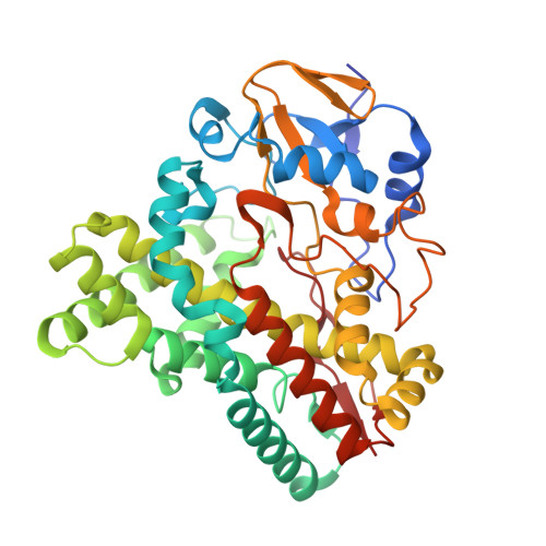
Crystal Structure of Cytochrome P450revI


Experimental Data Snapshot

  • Method: X-RAY DIFFRACTION
  • Resolution: 1.40 Å
  • R-Value Free: 
    0.188 (Depositor), 0.187 (DCC) 
  • R-Value Work: 
    0.173 (Depositor), 0.172 (DCC) 
  • R-Value Observed: 
    0.174 (Depositor) 

wwPDB Validation 3D Report Full Report

Validation slider image for 3WVS

Ligand Structure Quality Assessment 


This is version 1.5 of the entry. See complete history. 

Literature

Structure-function analyses of cytochrome P450revI involved in reveromycin A biosynthesis and evaluation of the biological activity of its substrate, reveromycin T.

Takahashi, S., Nagano, S., Nogawa, T., Kanoh, N., Uramoto, M., Kawatani, M., Shimizu, T., Miyazawa, T., Shiro, Y., Osada, H.

(2014) J Biol Chem 289: 32446-32458

  • DOI: https://doi.org/10.1074/jbc.M114.598391
  • Primary Citation Related Structures: 
    3WVS

  • PubMed Abstract: 

    Numerous cytochrome P450s are involved in secondary metabolite biosynthesis. The biosynthetic gene cluster for reveromycin A (RM-A), which is a promising lead compound with anti-osteoclastic activity, also includes a P450 gene, revI. To understand the roles of P450revI, we comprehensively characterized the enzyme by genetic, kinetic, and structural studies. The revI gene disruptants (ΔrevI) resulted in accumulation of reveromycin T (RM-T), and revI gene complementation restored RM-A production, indicating that the physiological substrate of P450revI is RM-T. Indeed, the purified P450revI catalyzed the C18-hydroxylation of RM-T more efficiently than the other RM derivatives tested. Moreover, the 1.4 Å resolution co-crystal structure of P450revI with RM-T revealed that the substrate binds the enzyme with a folded compact conformation for C18-hydroxylation. To address the structure-enzyme activity relationship, site-directed mutagenesis was performed in P450revI. R190A and R81A mutations, which abolished salt bridge formation with C1 and C24 carboxyl groups of RM-T, respectively, resulted in significant loss of enzyme activity. The interaction between Arg(190) and the C1 carboxyl group of RM-T elucidated why P450revI was unable to catalyze both RM-T 1-methyl ester and RM-T 1-ethyl ester. Moreover, the accumulation of RM-T in ΔrevI mutants enabled us to characterize its biological activity. Our results show that RM-T had stronger anticancer activity and isoleucyl-tRNA synthetase inhibition than RM-A. However, RM-T showed much less anti-osteoclastic activity than RM-A, indicating that hemisuccinate moiety is important for the activity. Structure-based P450revI engineering for novel hydroxylation and subsequent hemisuccinylation will help facilitate the development of RM derivatives with anti-osteoclast activity.


  • Organizational Affiliation: 
    • From the Chemical Biology Group, RIKEN Center for Sustainable Resource Science, Saitama 351-0198, Japan, the Antibiotics Laboratory, RIKEN, Saitama 351-0198, Japan, shunjitaka@riken.jp.

Macromolecule Content 

  • Total Structure Weight: 46.52 kDa 
  • Atom Count: 3,699 
  • Modeled Residue Count: 393 
  • Deposited Residue Count: 401 
  • Unique protein chains: 1

Macromolecules

Find similar proteins by:|  3D Structure
Entity ID: 1
MoleculeChains  Sequence LengthOrganismDetailsImage
Putative monooxygenase401Actinacidiphila reveromycinicaMutation(s): 0 
Gene Names: revI
UniProt
Find proteins for G1UDU7 (Actinacidiphila reveromycinica)
Explore G1UDU7 
Go to UniProtKB:  G1UDU7
Entity Groups
Sequence Clusters30% Identity50% Identity70% Identity90% Identity95% Identity100% Identity
UniProt GroupG1UDU7
Sequence Annotations
Expand
Reference Sequence

Small Molecules

Ligands 4 Unique
IDChains Name / Formula / InChI Key2D Diagram3D Interactions
HEM

Query on HEM


Download:Ideal Coordinates CCD File
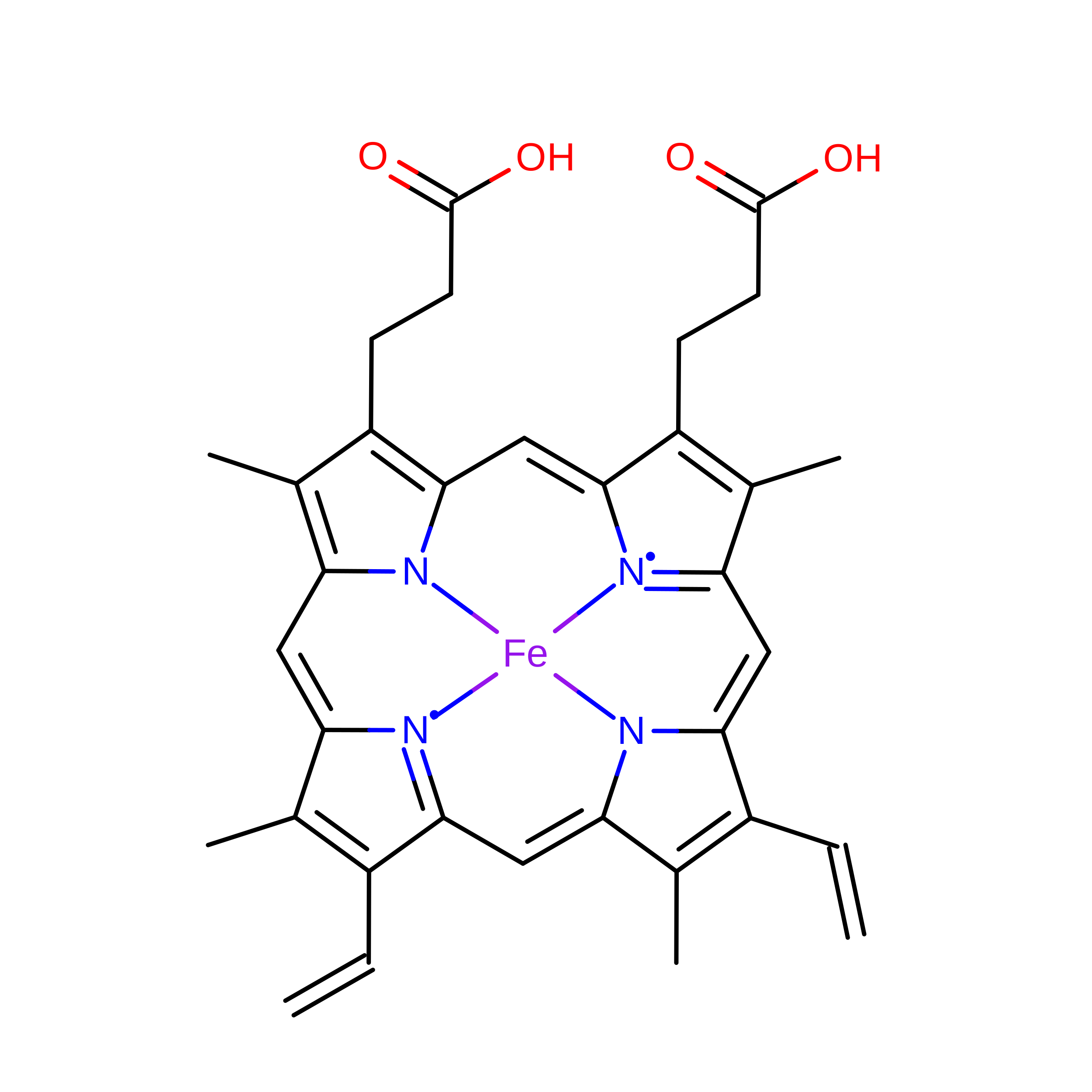
B [auth A]PROTOPORPHYRIN IX CONTAINING FE
C34 H32 Fe N4 O4
KABFMIBPWCXCRK-RGGAHWMASA-L
RRM

Query on RRM


Download:Ideal Coordinates CCD File
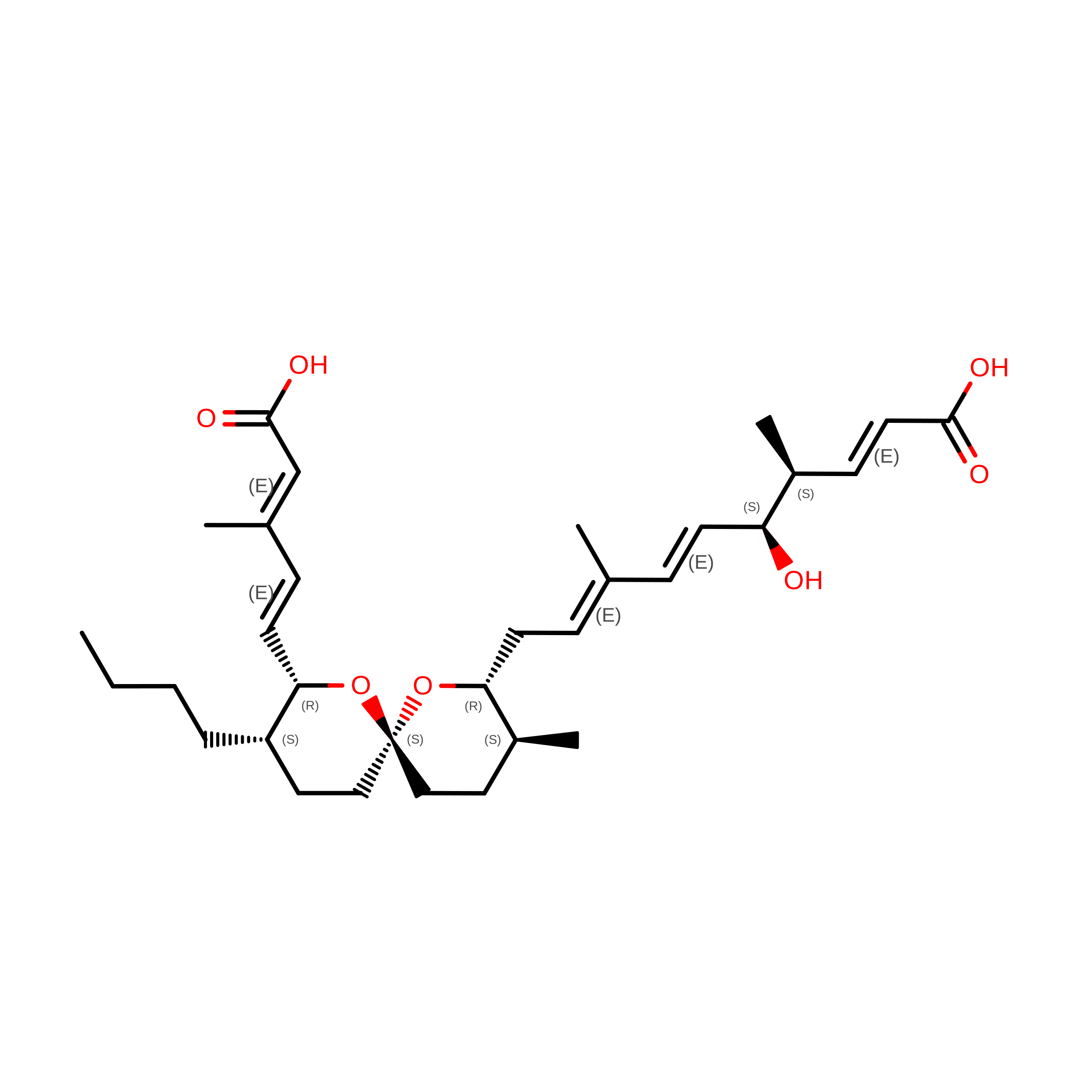
C [auth A](2E,4S,5S,6E,8E)-10-{(2R,3S,6S,8R,9S)-9-butyl-8-[(1E,3E)-4-carboxy-3-methylbuta-1,3-dien-1-yl]-3-methyl-1,7-dioxaspiro[5.5]undec-2-yl}-5-hydroxy-4,8-dimethyldeca-2,6,8-trienoic acid
C32 H48 O7
YEZCSKCHOPBNCC-OXNFKLAGSA-N
TLA

Query on TLA


Download:Ideal Coordinates CCD File
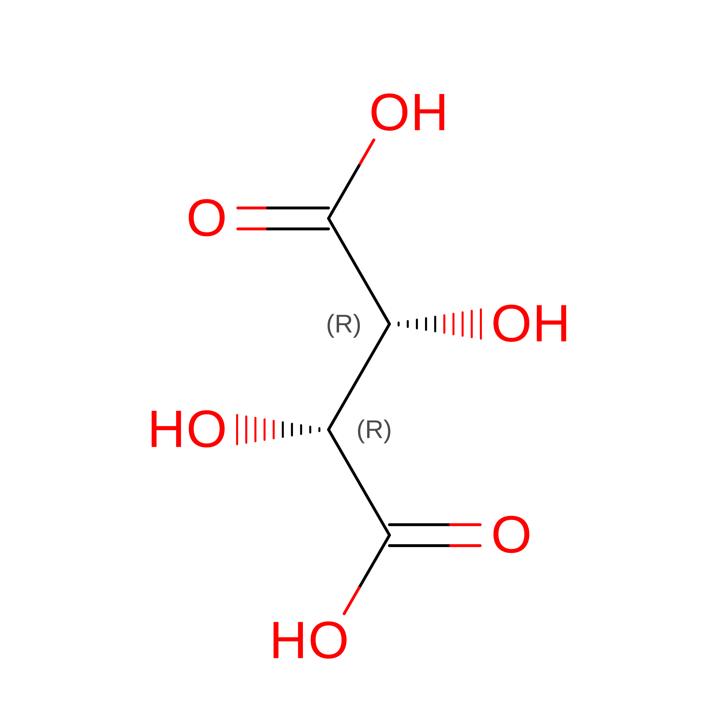
D [auth A]L(+)-TARTARIC ACID
C4 H6 O6
FEWJPZIEWOKRBE-JCYAYHJZSA-N
GOL

Query on GOL


Download:Ideal Coordinates CCD File
E [auth A],
F [auth A],
G [auth A],
H [auth A],
I [auth A]
GLYCEROL
C3 H8 O3
PEDCQBHIVMGVHV-UHFFFAOYSA-N

Experimental Data & Validation

Experimental Data

  • Method: X-RAY DIFFRACTION
  • Resolution: 1.40 Å
  • R-Value Free:  0.188 (Depositor), 0.187 (DCC) 
  • R-Value Work:  0.173 (Depositor), 0.172 (DCC) 
  • R-Value Observed: 0.174 (Depositor) 
Space Group: P 1 21 1
Unit Cell:
Length ( Å )Angle ( ˚ )
a = 52.779α = 90
b = 73.472β = 112.11
c = 58.305γ = 90
Software Package:
Software NamePurpose
DENZOdata reduction
SCALEPACKdata scaling
SOLVEphasing
RESOLVEphasing
REFMACrefinement
PDB_EXTRACTdata extraction
HKL-2000data collection
HKL-2000data reduction
HKL-2000data scaling

Structure Validation

View Full Validation Report


Ligand Structure Quality Assessment 


Entry History 

Deposition Data

Revision History  (Full details and data files)

  • Version 1.0: 2014-10-01
    Type: Initial release
  • Version 1.1: 2014-10-08
    Changes: Database references
  • Version 1.2: 2017-11-22
    Changes: Refinement description
  • Version 1.3: 2022-08-24
    Changes: Database references, Derived calculations
  • Version 1.4: 2024-05-29
    Changes: Data collection
  • Version 1.5: 2026-08-12
    Type: Remediation
    Reason: Metalloprotein remediation
    Changes: Derived calculations, Structure summary