3WLE | pdb_00003wle

Crystal structure of (R)-carbonyl reductase from Candida Parapsilosis in complex with NAD


Experimental Data Snapshot

  • Method: X-RAY DIFFRACTION
  • Resolution: 2.16 Å
  • R-Value Free: 
    0.216 (Depositor), 0.216 (DCC) 
  • R-Value Work: 
    0.181 (Depositor), 0.181 (DCC) 
  • R-Value Observed: 
    0.183 (Depositor) 

Starting Model: experimental
View more details

wwPDB Validation 3D Report Full Report

Validation slider image for 3WLE

Ligand Structure Quality Assessment 


This is version 1.2 of the entry. See complete history

Literature

Unconserved substrate-binding sites direct the stereoselectivity of medium-chain alcohol dehydrogenase

Wang, S.S.Nie, Y.Xu, Y.Zhang, R.Z.Ko, T.P.Huang, C.H.Chan, H.C.Guo, R.T.Xiao, R.

(2014) Chem Commun (Camb) 50: 7770-7772

  • DOI: https://doi.org/10.1039/c4cc01752h
  • Primary Citation Related Structures: 
    3WLE, 3WLF, 3WNQ

  • PubMed Abstract: 

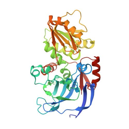
    Structure-guided design of substrate-binding pocket inversed the stereoselectivity of an NADH-dependent medium-chain alcohol dehydrogenase (MDR) from Prelog to anti-Prelog. The pocket-forming amino acids, especially the unconserved residues as hotspots, play critical roles in directing MDRs' stereoselectivity.


  • Organizational Affiliation
    • School of Biotechnology and Key Laboratory of Industrial Biotechnology, Ministry of Education, Jiangnan University, Wuxi 214122, China. yxu@jiangnan.edu.cn.

Macromolecule Content 

  • Total Structure Weight: 148.56 kDa 
  • Atom Count: 11,345 
  • Modeled Residue Count: 1,340 
  • Deposited Residue Count: 1,364 
  • Unique protein chains: 1

Macromolecules

Find similar proteins by:|  3D Structure
Entity ID: 1
MoleculeChains  Sequence LengthOrganismDetailsImage
(R)-specific carbonyl reductase
A, B, C, D
341Candida parapsilosisMutation(s): 0 
Gene Names: CPRADH
EC: 1.1.1.1
UniProt
Find proteins for A1X808 (Candida parapsilosis)
Explore A1X808 
Go to UniProtKB:  A1X808
Entity Groups
Sequence Clusters30% Identity50% Identity70% Identity90% Identity95% Identity100% Identity
UniProt GroupA1X808
Sequence Annotations
Expand
Reference Sequence

Small Molecules

Ligands 2 Unique
IDChains Name / Formula / InChI Key2D Diagram3D Interactions
NAD

Query on NAD


Download:Ideal Coordinates CCD File
E [auth A],
H [auth B],
K [auth C],
N [auth D]
NICOTINAMIDE-ADENINE-DINUCLEOTIDE
C21 H27 N7 O14 P2
BAWFJGJZGIEFAR-NNYOXOHSSA-N
ZN

Query on ZN


Download:Ideal Coordinates CCD File
F [auth A]
G [auth A]
I [auth B]
J [auth B]
L [auth C]
F [auth A],
G [auth A],
I [auth B],
J [auth B],
L [auth C],
M [auth C],
O [auth D],
P [auth D]
ZINC ION
Zn
PTFCDOFLOPIGGS-UHFFFAOYSA-N

Experimental Data & Validation

Experimental Data

  • Method: X-RAY DIFFRACTION
  • Resolution: 2.16 Å
  • R-Value Free:  0.216 (Depositor), 0.216 (DCC) 
  • R-Value Work:  0.181 (Depositor), 0.181 (DCC) 
  • R-Value Observed: 0.183 (Depositor) 
Space Group: P 21 21 21
Unit Cell:
Length ( Å )Angle ( ˚ )
a = 85.641α = 90
b = 106.105β = 90
c = 145.55γ = 90
Software Package:
Software NamePurpose
Blu-Icedata collection
PHASERphasing
CNSrefinement
HKL-2000data reduction
HKL-2000data scaling

Structure Validation

View Full Validation Report


Ligand Structure Quality Assessment 


Entry History 

Deposition Data

Revision History  (Full details and data files)

  • Version 1.0: 2014-07-16
    Type: Initial release
  • Version 1.1: 2017-11-22
    Changes: Refinement description
  • Version 1.2: 2023-11-08
    Changes: Data collection, Database references, Derived calculations, Refinement description