3W6B | pdb_00003w6b

Crystal structure of catalytic domain of chitinase from Ralstonia sp. A-471


Experimental Data Snapshot

  • Method: X-RAY DIFFRACTION
  • Resolution: 1.90 Å
  • R-Value Free: 
    0.229 (Depositor), 0.232 (DCC) 
  • R-Value Work: 
    0.185 (Depositor), 0.187 (DCC) 
  • R-Value Observed: 
    0.188 (Depositor) 

wwPDB Validation 3D Report Full Report

Validation slider image for 3W6B

This is version 1.3 of the entry. See complete history

Literature

Crystal Structures of the Catalytic Domain of a Novel Glycohydrolase Family 23 Chitinase from Ralstonia sp. A-471 Reveals a Unique Arrangement of the Catalytic Residues for Inverting Chitin Hydrolysis

Arimori, T.Kawamoto, N.Shinya, S.Okazaki, N.Nakazawa, M.Miyatake, K.Fukamizo, T.Ueda, M.Tamada, T.

(2013) J Biological Chem 288: 18696-18706

  • DOI: https://doi.org/10.1074/jbc.M113.462135
  • Primary Citation Related Structures: 
    3W6B, 3W6C, 3W6D, 3W6E, 3W6F

  • PubMed Abstract: 

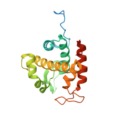
    Chitinase C from Ralstonia sp. A-471 (Ra-ChiC) has a catalytic domain sequence similar to goose-type (G-type) lysozymes and, unlike other chitinases, belongs to glycohydrolase (GH) family 23. Using NMR spectroscopy, however, Ra-ChiC was found to interact only with the chitin dimer but not with the peptidoglycan fragment. Here we report the crystal structures of wild-type, E141Q, and E162Q of the catalytic domain of Ra-ChiC with or without chitin oligosaccharides. Ra-ChiC has a substrate-binding site including a tunnel-shaped cavity, which determines the substrate specificity. Mutation analyses based on this structural information indicated that a highly conserved Glu-141 acts as a catalytic acid, and that Asp-226 located at the roof of the tunnel activates a water molecule as a catalytic base. The unique arrangement of the catalytic residues makes a clear contrast to the other GH23 members and also to inverting GH19 chitinases.


  • Organizational Affiliation
    • Quantum Beam Science Directorate, Japan Atomic Energy Agency, 2-4 Shirakata-Shirane, Tokai, Ibaraki 319-1195, Japan.

Macromolecule Content 

  • Total Structure Weight: 81.71 kDa 
  • Atom Count: 5,094 
  • Modeled Residue Count: 619 
  • Deposited Residue Count: 732 
  • Unique protein chains: 1

Macromolecules

Find similar proteins by:|  3D Structure
Entity ID: 1
MoleculeChains  Sequence LengthOrganismDetailsImage
Lysozyme-like chitinolytic enzyme
A, B, C, D
183Ralstonia sp. A-471Mutation(s): 0 
EC: 3.2.1.14
UniProt
Find proteins for B7XCV4 (Ralstonia sp. A-471)
Explore B7XCV4 
Go to UniProtKB:  B7XCV4
Entity Groups
Sequence Clusters30% Identity50% Identity70% Identity90% Identity95% Identity100% Identity
UniProt GroupB7XCV4
Sequence Annotations
Expand
Reference Sequence

Experimental Data & Validation

Experimental Data

  • Method: X-RAY DIFFRACTION
  • Resolution: 1.90 Å
  • R-Value Free:  0.229 (Depositor), 0.232 (DCC) 
  • R-Value Work:  0.185 (Depositor), 0.187 (DCC) 
  • R-Value Observed: 0.188 (Depositor) 
Space Group: P 61 2 2
Unit Cell:
Length ( Å )Angle ( ˚ )
a = 99.66α = 90
b = 99.66β = 90
c = 242.464γ = 120
Software Package:
Software NamePurpose
DENZOdata reduction
SCALEPACKdata scaling
REFMACrefinement
PDB_EXTRACTdata extraction
HKL-2000data collection
HKL-2000data reduction
HKL-2000data scaling
SOLVEphasing

Structure Validation

View Full Validation Report


Entry History 

Deposition Data

Revision History  (Full details and data files)

  • Version 1.0: 2013-05-15
    Type: Initial release
  • Version 1.1: 2013-08-14
    Changes: Database references
  • Version 1.2: 2017-11-22
    Changes: Refinement description
  • Version 1.3: 2024-03-20
    Changes: Data collection, Database references, Derived calculations