3VSJ | pdb_00003vsj

Crystal structure of 1,6-APD (2-ANIMOPHENOL-1,6-DIOXYGENASE) complexed with intermediate products


Experimental Data Snapshot

  • Method: X-RAY DIFFRACTION
  • Resolution: 2.30 Å
  • R-Value Free: 
    0.234 (Depositor), 0.235 (DCC) 
  • R-Value Work: 
    0.190 (Depositor), 0.191 (DCC) 
  • R-Value Observed: 
    0.192 (Depositor) 

Starting Model: experimental
View more details

wwPDB Validation 3D Report Full Report

Validation slider image for 3VSJ

This is version 1.1 of the entry. See complete history

Literature

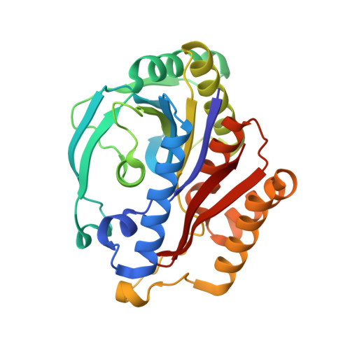
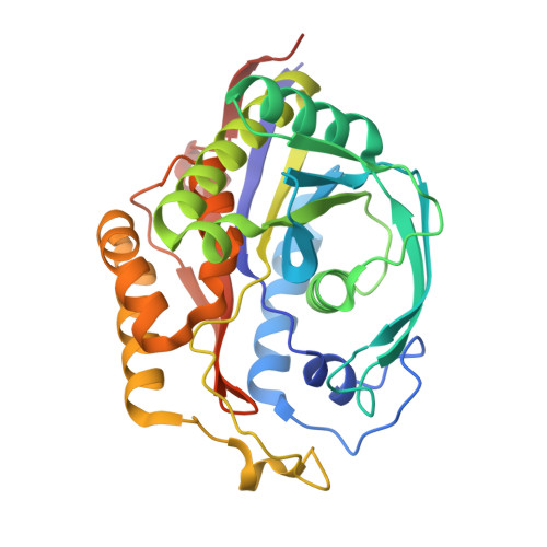
Structures of aminophenol dioxygenase in complex with intermediate, product and inhibitor

Li, D.F.Zhang, J.Y.Hou, Y.J.Liu, L.Hu, Y.Liu, S.J.Wang, D.C.Liu, W.

(2013) Acta Crystallogr D Biol Crystallogr 69: 32-43

  • DOI: https://doi.org/10.1107/S0907444912042072
  • Primary Citation Related Structures: 
    3VSG, 3VSH, 3VSI, 3VSJ

  • PubMed Abstract: 

    Dioxygen activation by nonhaem Fe(II) enzymes containing the 2-His-1-carboxylate facial triad has been extensively studied in recent years. Here, crystal structures of 2-aminophenol 1,6-dioxygenase, an enzyme that represents a minor group of extradiol dioxygenases and that catalyses the ring opening of 2-aminophenol, in complex with the lactone intermediate (4Z,6Z)-3-iminooxepin-2(3H)-one and the product 2-aminomuconic 6-semialdehyde and in complex with the suicide inhibitor 4-nitrocatechol are reported. The Fe-ligand binding schemes observed in these structures revealed some common geometrical characteristics that are shared by the published structures of extradiol dioxygenases, suggesting that enzymes that catalyse the oxidation of noncatecholic compounds are very likely to utilize a similar strategy for dioxygen activation and the fission of aromatic rings as the canonical mechanism. The Fe-ligation arrangement, however, is strikingly enantiomeric to that of all other 2-His-1-carboxylate enzymes apart from protocatechuate 4,5-dioxygenase. This structural variance leads to the generation of an uncommon O(-)-Fe(2+)-O(-) species prior to O(2) binding, which probably forms the structural basis on which APD distinguishes its specific substrate and inhibitor, which share an analogous molecular structure.


  • Organizational Affiliation
    • National Laboratory of Biomacromolecules, Institute of Biophysics, Chinese Academy of Sciences, Beijing, People's Republic of China.

Macromolecule Content 

  • Total Structure Weight: 129.19 kDa 
  • Atom Count: 9,950 
  • Modeled Residue Count: 1,142 
  • Deposited Residue Count: 1,166 
  • Unique protein chains: 2

Macromolecules

Find similar proteins by:|  3D Structure
Entity ID: 1
MoleculeChains  Sequence LengthOrganismDetailsImage
2-amino-5-chlorophenol 1,6-dioxygenase alpha subunit
A, C
271Comamonas testosteroni CNB-1Mutation(s): 0 
EC: 1.13.11.8
UniProt
Find proteins for Q6J1Z5 (Comamonas testosteroni)
Explore Q6J1Z5 
Go to UniProtKB:  Q6J1Z5
Entity Groups
Sequence Clusters30% Identity50% Identity70% Identity90% Identity95% Identity100% Identity
UniProt GroupQ6J1Z5
Sequence Annotations
Expand
Reference Sequence
Find similar proteins by:|  3D Structure
Entity ID: 2
MoleculeChains  Sequence LengthOrganismDetailsImage
2-amino-5-chlorophenol 1,6-dioxygenase beta subunit
B, D
312Comamonas testosteroni CNB-1Mutation(s): 0 
EC: 1.13.11.8 (PDB Primary Data), 1.13.11.76 (UniProt), 1.13.11.74 (UniProt)
UniProt
Find proteins for Q6J1Z6 (Comamonas testosteroni)
Explore Q6J1Z6 
Go to UniProtKB:  Q6J1Z6
Entity Groups
Sequence Clusters30% Identity50% Identity70% Identity90% Identity95% Identity100% Identity
UniProt GroupQ6J1Z6
Sequence Annotations
Expand
Reference Sequence

Small Molecules

Ligands 5 Unique
IDChains Name / Formula / InChI Key2D Diagram3D Interactions
2XP

Query on 2XP


Download:Ideal Coordinates CCD File
G [auth B](2Z,4Z)-2-imino-6-oxohex-4-enoic acid
C6 H7 N O3
IZBQCPKKCVTOTK-PQZOIKATSA-N
2X7

Query on 2X7


Download:Ideal Coordinates CCD File
I [auth D](3E)-3-iminooxepin-2(3H)-one
C6 H5 N O2
HQPIXVSGYRVKOL-FNORWQNLSA-N
FE2

Query on FE2


Download:Ideal Coordinates CCD File
F [auth B],
H [auth D]
FE (II) ION
Fe
CWYNVVGOOAEACU-UHFFFAOYSA-N
CL

Query on CL


Download:Ideal Coordinates CCD File
E [auth A]CHLORIDE ION
Cl
VEXZGXHMUGYJMC-UHFFFAOYSA-M
OH

Query on OH


Download:Ideal Coordinates CCD File
J [auth D]HYDROXIDE ION
H O
XLYOFNOQVPJJNP-UHFFFAOYSA-M

Experimental Data & Validation

Experimental Data

  • Method: X-RAY DIFFRACTION
  • Resolution: 2.30 Å
  • R-Value Free:  0.234 (Depositor), 0.235 (DCC) 
  • R-Value Work:  0.190 (Depositor), 0.191 (DCC) 
  • R-Value Observed: 0.192 (Depositor) 
Space Group: C 1 2 1
Unit Cell:
Length ( Å )Angle ( ˚ )
a = 270.24α = 90
b = 48.39β = 109.57
c = 108.55γ = 90
Software Package:
Software NamePurpose
CrystalCleardata collection
PHASESphasing
REFMACrefinement
MOSFLMdata reduction
SCALAdata scaling

Structure Validation

View Full Validation Report


Entry History 

Deposition Data

Revision History  (Full details and data files)

  • Version 1.0: 2013-01-16
    Type: Initial release
  • Version 1.1: 2023-11-08
    Changes: Data collection, Database references, Derived calculations, Refinement description