Structural elucidation of dextran degradation mechanism by streptococcus mutans dextranase belonging to glycoside hydrolase family 66
Suzuki, N., Kim, Y.M., Fujimoto, Z., Momma, M., Okuyama, M., Mori, H., Funane, K., Kimura, A.(2012) J Biol Chem 287: 19916-19926
- PubMed: 22337884 Search on PubMedSearch on PubMed Central
- DOI: https://doi.org/10.1074/jbc.M112.342444
- Primary Citation Related Structures:
3VMN, 3VMO, 3VMP - PubMed Abstract:
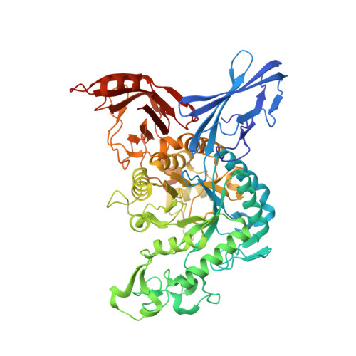
Dextranase is an enzyme that hydrolyzes dextran α-1,6 linkages. Streptococcus mutans dextranase belongs to glycoside hydrolase family 66, producing isomaltooligosaccharides of various sizes and consisting of at least five amino acid sequence regions. The crystal structure of the conserved fragment from Gln(100) to Ile(732) of S. mutans dextranase, devoid of its N- and C-terminal variable regions, was determined at 1.6 Å resolution and found to contain three structural domains. Domain N possessed an immunoglobulin-like β-sandwich fold; domain A contained the enzyme's catalytic module, comprising a (β/α)(8)-barrel; and domain C formed a β-sandwich structure containing two Greek key motifs. Two ligand complex structures were also determined, and, in the enzyme-isomaltotriose complex structure, the bound isomaltooligosaccharide with four glucose moieties was observed in the catalytic glycone cleft and considered to be the transglycosylation product of the enzyme, indicating the presence of four subsites, -4 to -1, in the catalytic cleft. The complexed structure with 4',5'-epoxypentyl-α-d-glucopyranoside, a suicide substrate of the enzyme, revealed that the epoxide ring reacted to form a covalent bond with the Asp(385) side chain. These structures collectively indicated that Asp(385) was the catalytic nucleophile and that Glu(453) was the acid/base of the double displacement mechanism, in which the enzyme showed a retaining catalytic character. This is the first structural report for the enzyme belonging to glycoside hydrolase family 66, elucidating the enzyme's catalytic machinery.
- Biomolecular Research Unit, National Institute of Agrobiological Sciences, 2-1-2 Kannondai, Tsukuba 305-8602, Japan.
Organizational Affiliation: