3UOR | pdb_00003uor

The structure of the sugar-binding protein MalE from the phytopathogen Xanthomonas citri


Experimental Data Snapshot

  • Method: X-RAY DIFFRACTION
  • Resolution: 2.20 Å
  • R-Value Free: 
    0.228 (Depositor), 0.221 (DCC) 
  • R-Value Work: 
    0.189 (Depositor), 0.184 (DCC) 
  • R-Value Observed: 
    0.191 (Depositor) 

wwPDB Validation 3D Report Full Report

Validation slider image for 3UOR

This is version 1.4 of the entry. See complete history

Literature

Structure determination of a sugar-binding protein from the phytopathogenic bacterium Xanthomonas citri.

Medrano, F.J.de Souza, C.S.Romero, A.Balan, A.

(2014) Acta Crystallogr Sect F Struct Biol Cryst Commun 70: 564-571

  • DOI: https://doi.org/10.1107/S2053230X14006578
  • Primary Citation Related Structures: 
    3UOR

  • PubMed Abstract: 

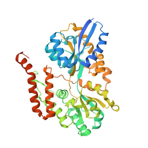
    The uptake of maltose and related sugars in Gram-negative bacteria is mediated by an ABC transporter encompassing a periplasmic component (the maltose-binding protein or MalE), a pore-forming membrane protein (MalF and MalG) and a membrane-associated ATPase (MalK). In the present study, the structure determination of the apo form of the putative maltose/trehalose-binding protein (Xac-MalE) from the citrus pathogen Xanthomonas citri in space group P6522 is described. The crystals contained two protein molecules in the asymmetric unit and diffracted to 2.8 Å resolution. Xac-MalE conserves the structural and functional features of sugar-binding proteins and a ligand-binding pocket with similar characteristics to eight different orthologues, including the residues for maltose and trehalose interaction. This is the first structure of a sugar-binding protein from a phytopathogenic bacterium, which is highly conserved in all species from the Xanthomonas genus.


  • Organizational Affiliation
    • Department of Chemical and Physical Biology, Centro de Investigaciones Biologicas (CSIC), Madrid, Spain.

Macromolecule Content 

  • Total Structure Weight: 102.77 kDa 
  • Atom Count: 6,719 
  • Modeled Residue Count: 792 
  • Deposited Residue Count: 916 
  • Unique protein chains: 1

Macromolecules

Find similar proteins by:|  3D Structure
Entity ID: 1
MoleculeChains  Sequence LengthOrganismDetailsImage
ABC transporter sugar binding protein
A, B
458Xanthomonas citri subsp. citri 306Mutation(s): 0 
Gene Names: malEXAC2310

Experimental Data & Validation

Experimental Data

  • Method: X-RAY DIFFRACTION
  • Resolution: 2.20 Å
  • R-Value Free:  0.228 (Depositor), 0.221 (DCC) 
  • R-Value Work:  0.189 (Depositor), 0.184 (DCC) 
  • R-Value Observed: 0.191 (Depositor) 
Space Group: P 65 2 2
Unit Cell:
Length ( Å )Angle ( ˚ )
a = 122.971α = 90
b = 122.971β = 90
c = 304.28γ = 120
Software Package:
Software NamePurpose
MxCuBEdata collection
SHARPphasing
PHENIXrefinement
XDSdata reduction
SCALAdata scaling

Structure Validation

View Full Validation Report


Entry History 

Deposition Data

Revision History  (Full details and data files)

  • Version 1.0: 2011-12-14
    Type: Initial release
  • Version 1.1: 2014-05-14
    Changes: Database references
  • Version 1.2: 2014-09-24
    Changes: Database references
  • Version 1.3: 2017-11-08
    Changes: Refinement description
  • Version 1.4: 2024-02-28
    Changes: Data collection, Database references