3U1P | pdb_00003u1p

Crystal Structure of M. tuberculosis LD-transpeptidase type 2 with Modified Catalytic Cysteine (C354)


Experimental Data Snapshot

  • Method: X-RAY DIFFRACTION
  • Resolution: 2.80 Å
  • R-Value Free: 
    0.258 (Depositor), 0.252 (DCC) 
  • R-Value Work: 
    0.187 (Depositor), 0.182 (DCC) 
  • R-Value Observed: 
    0.190 (Depositor) 

wwPDB Validation 3D Report Full Report

Validation slider image for 3U1P

This is version 1.4 of the entry. See complete history

Literature

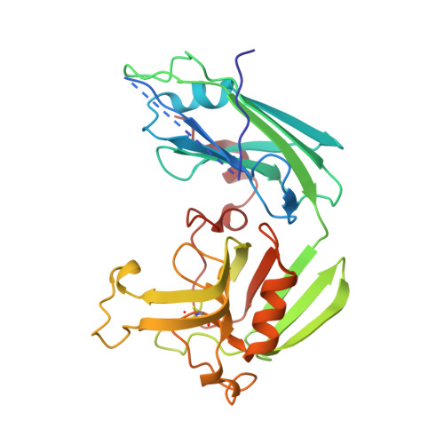
Targeting the Cell Wall of Mycobacterium tuberculosis: Structure and Mechanism of L,D-Transpeptidase 2.

Erdemli, S.B.Gupta, R.Bishai, W.R.Lamichhane, G.Amzel, L.M.Bianchet, M.A.

(2012) Structure 20: 2103-2115

  • DOI: https://doi.org/10.1016/j.str.2012.09.016
  • Primary Citation Related Structures: 
    3TUR, 3TX4, 3U1P, 3VAE

  • PubMed Abstract: 

    With multidrug-resistant cases of tuberculosis increasing globally, better antibiotic drugs and novel drug targets are becoming an urgent need. Traditional β-lactam antibiotics that inhibit D,D-transpeptidases are not effective against mycobacteria, in part because mycobacteria rely mostly on L,D-transpeptidases for biosynthesis and maintenance of their peptidoglycan layer. This reliance plays a major role in drug resistance and persistence of Mycobacterium tuberculosis (Mtb) infections. The crystal structure at 1.7 Å resolution of the Mtb L,D-transpeptidase Ldt(Mt2) containing a bound peptidoglycan fragment, reported here, provides information about catalytic site organization as well as substrate recognition by the enzyme. Based on our structural, kinetic, and calorimetric data, we propose a catalytic mechanism for Ldt(Mt2) in which both acyl-acceptor and acyl-donor substrates reach the catalytic site from the same, rather than different, entrances. Together, this information provides vital insights to facilitate development of drugs targeting this validated yet unexploited enzyme.


  • Organizational Affiliation
    • Department of Biophysics and Biophysical Chemistry, Johns Hopkins University School of Medicine, Baltimore, MD 21205, USA.

Macromolecule Content 

  • Total Structure Weight: 62.5 kDa 
  • Atom Count: 4,244 
  • Modeled Residue Count: 524 
  • Deposited Residue Count: 574 
  • Unique protein chains: 1

Macromolecules

Find similar proteins by:|  3D Structure
Entity ID: 1
MoleculeChains  Sequence LengthOrganismDetailsImage
Mycobacteria Tuberculosis LD-transpeptidase type 2
A, B
287Mycobacterium tuberculosisMutation(s): 0 
Gene Names: lppSMT2594Rv2518c
EC: 2.3.2
UniProt
Find proteins for O53223 (Mycobacterium tuberculosis (strain CDC 1551 / Oshkosh))
Explore O53223 
Go to UniProtKB:  O53223
Entity Groups
Sequence Clusters30% Identity50% Identity70% Identity90% Identity95% Identity100% Identity
UniProt GroupO53223
Sequence Annotations
Expand
Reference Sequence

Small Molecules

Modified Residues  1 Unique
IDChains TypeFormula2D DiagramParent
CSO
Query on CSO
A, B
L-PEPTIDE LINKINGC3 H7 N O3 SCYS

Experimental Data & Validation

Experimental Data

  • Method: X-RAY DIFFRACTION
  • Resolution: 2.80 Å
  • R-Value Free:  0.258 (Depositor), 0.252 (DCC) 
  • R-Value Work:  0.187 (Depositor), 0.182 (DCC) 
  • R-Value Observed: 0.190 (Depositor) 
Space Group: I 21 21 21
Unit Cell:
Length ( Å )Angle ( ˚ )
a = 119.044α = 90
b = 121.223β = 90
c = 122.884γ = 90
Software Package:
Software NamePurpose
SCALEPACKdata scaling
REFMACrefinement
PDB_EXTRACTdata extraction
SHARPphasing

Structure Validation

View Full Validation Report


Entry History 

Deposition Data

Revision History  (Full details and data files)

  • Version 1.0: 2012-12-05
    Type: Initial release
  • Version 1.1: 2012-12-12
    Changes: Database references
  • Version 1.2: 2012-12-26
    Changes: Database references
  • Version 1.3: 2018-04-04
    Changes: Data collection
  • Version 1.4: 2024-10-16
    Changes: Data collection, Database references, Derived calculations, Structure summary