The X-ray crystal structure of a pseudoazurin from Sinorhizobium meliloti.
Laming, E.M., McGrath, A.P., Guss, J.M., Kappler, U., Maher, M.J.(2012) J Inorg Biochem 115: 148-154
- PubMed: 22776735 Search on PubMed
- DOI: https://doi.org/10.1016/j.jinorgbio.2012.04.005
- Primary Citation Related Structures:
3TU6 - PubMed Abstract:
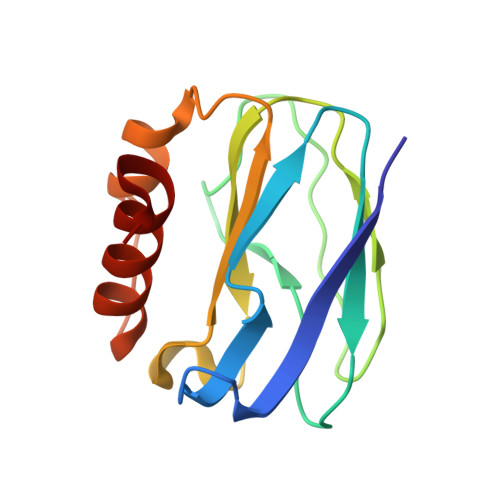
The X-ray crystal structure of oxidised pseudoazurin from the denitrifying plant symbiotic bacterium Sinorhizobium meliloti (SmPAz2) has been solved to a resolution of 2.0 Å. The pseudoazurin from Sinorhizobium sp. is unusual as it forms an operon with a sulfite dehydrogenase enzyme, rather than a Cu nitrite reductase. Examination of the structure reveals that the geometric parameters of the Type I Cu site in SmPAz2 correlate with observed features in the electronic spectrum of the protein. Comparison of the structure of SmPAz2 with those of pseudoazurins from five other bacterial species shows that the surface of SmPAz2 bears a conserved hydrophobic patch encircled by positively-charged residues, which may serve as a recognition site for its redox partners.
- School of Molecular Bioscience, University of Sydney, NSW 2006, Australia.
Organizational Affiliation: