3T7H | pdb_00003t7h

Atg8 transfer from Atg7 to Atg3: a distinctive E1-E2 architecture and mechanism in the autophagy pathway


Experimental Data Snapshot

  • Method: X-RAY DIFFRACTION
  • Resolution: 1.60 Å
  • R-Value Free: 
    0.206 (Depositor), 0.211 (DCC) 
  • R-Value Work: 
    0.186 (Depositor) 
  • R-Value Observed: 
    0.187 (Depositor) 

Starting Model: experimental
View more details

wwPDB Validation 3D Report Full Report

Validation slider image for 3T7H

This is version 1.1 of the entry. See complete history

Literature

Atg8 transfer from atg7 to atg3: a distinctive e1-e2 architecture and mechanism in the autophagy pathway.

Taherbhoy, A.M.Tait, S.W.Kaiser, S.E.Williams, A.H.Deng, A.Nourse, A.Hammel, M.Kurinov, I.Rock, C.O.Green, D.R.Schulman, B.A.

(2011) Mol Cell 44: 451-461

  • DOI: https://doi.org/10.1016/j.molcel.2011.08.034
  • Primary Citation Related Structures: 
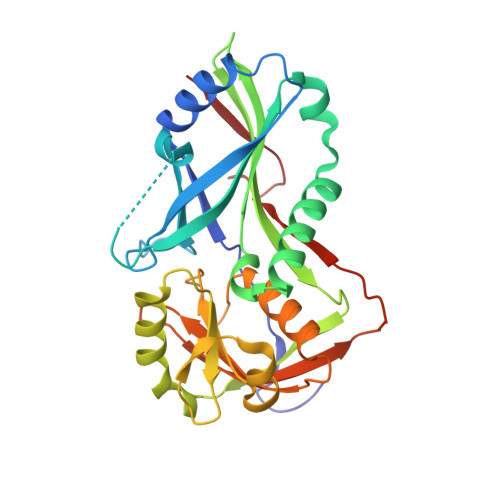
    3T7E, 3T7F, 3T7G, 3T7H

  • PubMed Abstract: 

    Atg7 is a noncanonical, homodimeric E1 enzyme that interacts with the noncanonical E2 enzyme, Atg3, to mediate conjugation of the ubiquitin-like protein (UBL) Atg8 during autophagy. Here we report that the unique N-terminal domain of Atg7 (Atg7(NTD)) recruits a unique "flexible region" from Atg3 (Atg3(FR)). The structure of an Atg7(NTD)-Atg3(FR) complex reveals hydrophobic residues from Atg3 engaging a conserved groove in Atg7, important for Atg8 conjugation. We also report the structure of the homodimeric Atg7 C-terminal domain, which is homologous to canonical E1s and bacterial antecedents. The structures, SAXS, and crosslinking data allow modeling of a full-length, dimeric (Atg7~Atg8-Atg3)(2) complex. The model and biochemical data provide a rationale for Atg7 dimerization: Atg8 is transferred in trans from the catalytic cysteine of one Atg7 protomer to Atg3 bound to the N-terminal domain of the opposite Atg7 protomer within the homodimer. The studies reveal a distinctive E1~UBL-E2 architecture for enzymes mediating autophagy.


  • Organizational Affiliation
    • Department of Structural Biology, St. Jude Children's Research Hospital, Memphis, TN 38105, USA.

Macromolecule Content 

  • Total Structure Weight: 66.88 kDa 
  • Atom Count: 5,325 
  • Modeled Residue Count: 568 
  • Deposited Residue Count: 582 
  • Unique protein chains: 1

Macromolecules

Find similar proteins by:|  3D Structure
Entity ID: 1
MoleculeChains  Sequence LengthOrganismDetailsImage
Ubiquitin-like modifier-activating enzyme ATG7
A, B
291Saccharomyces cerevisiaeMutation(s): 1 
Gene Names: ATG7APG7CVT2YHR171W
UniProt
Find proteins for P38862 (Saccharomyces cerevisiae (strain ATCC 204508 / S288c))
Explore P38862 
Go to UniProtKB:  P38862
Entity Groups
Sequence Clusters30% Identity50% Identity70% Identity90% Identity95% Identity100% Identity
UniProt GroupP38862
Sequence Annotations
Expand
Reference Sequence

Experimental Data & Validation

Experimental Data

  • Method: X-RAY DIFFRACTION
  • Resolution: 1.60 Å
  • R-Value Free:  0.206 (Depositor), 0.211 (DCC) 
  • R-Value Work:  0.186 (Depositor) 
  • R-Value Observed: 0.187 (Depositor) 
Space Group: P 21 21 21
Unit Cell:
Length ( Å )Angle ( ˚ )
a = 56.184α = 90
b = 74.439β = 90
c = 153.373γ = 90
Software Package:
Software NamePurpose
PHENIXmodel building
PHENIXrefinement
HKL-2000data reduction
HKL-2000data scaling
PHENIXphasing

Structure Validation

View Full Validation Report


Entry History 

Deposition Data

Revision History  (Full details and data files)

  • Version 1.0: 2011-11-23
    Type: Initial release
  • Version 1.1: 2023-09-13
    Changes: Data collection, Database references, Refinement description