3SYQ

Crystal structure of the G protein-gated inward rectifier K+ channel GIRK2 (Kir3.2) R201A mutant in complex with PIP2


Experimental Data Snapshot

  • Method: X-RAY DIFFRACTION
  • Resolution: 3.44 Å
  • R-Value Free: 0.323 
  • R-Value Work: 0.299 
  • R-Value Observed: 0.301 

Starting Model: experimental
View more details

wwPDB Validation   3D Report Full Report


Ligand Structure Quality Assessment 


This is version 1.1 of the entry. See complete history


Literature

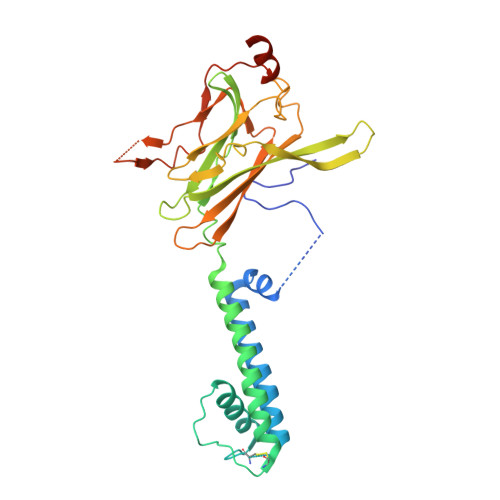
Crystal Structure of the Mammalian GIRK2 K(+) Channel and Gating Regulation by G Proteins, PIP(2), and Sodium.

Whorton, M.R.Mackinnon, R.

(2011) Cell 147: 199-208

  • DOI: https://doi.org/10.1016/j.cell.2011.07.046
  • Primary Citation of Related Structures:  
    3SYA, 3SYC, 3SYO, 3SYP, 3SYQ

  • PubMed Abstract: 

    G protein-gated K(+) channels (Kir3.1-Kir3.4) control electrical excitability in many different cells. Among their functions relevant to human physiology and disease, they regulate the heart rate and govern a wide range of neuronal activities. Here, we present the first crystal structures of a G protein-gated K(+) channel. By comparing the wild-type structure to that of a constitutively active mutant, we identify a global conformational change through which G proteins could open a G loop gate in the cytoplasmic domain. The structures of both channels in the absence and presence of PIP(2) suggest that G proteins open only the G loop gate in the absence of PIP(2), but in the presence of PIP(2) the G loop gate and a second inner helix gate become coupled, so that both gates open. We also identify a strategically located Na(+) ion-binding site, which would allow intracellular Na(+) to modulate GIRK channel activity. These data provide a structural basis for understanding multiligand regulation of GIRK channel gating.


  • Organizational Affiliation

    Laboratory of Molecular Neurobiology and Biophysics, Rockefeller University, New York, NY 10065, USA.


Macromolecules
Find similar proteins by:  (by identity cutoff)  |  3D Structure
Entity ID: 1
MoleculeChains Sequence LengthOrganismDetailsImage
G protein-activated inward rectifier potassium channel 2
A, B
340Mus musculusMutation(s): 1 
Gene Names: Girk2Kcnj6Kcnj7W
Membrane Entity: Yes 
UniProt & NIH Common Fund Data Resources
Find proteins for P48542 (Mus musculus)
Explore P48542 
Go to UniProtKB:  P48542
IMPC:  MGI:104781
Entity Groups  
Sequence Clusters30% Identity50% Identity70% Identity90% Identity95% Identity100% Identity
UniProt GroupP48542
Sequence Annotations
Expand
  • Reference Sequence
Experimental Data & Validation

Experimental Data

  • Method: X-RAY DIFFRACTION
  • Resolution: 3.44 Å
  • R-Value Free: 0.323 
  • R-Value Work: 0.299 
  • R-Value Observed: 0.301 
  • Space Group: C 2 2 21
Unit Cell:
Length ( Å )Angle ( ˚ )
a = 87.605α = 90
b = 208.491β = 90
c = 117.376γ = 90
Software Package:
Software NamePurpose
SCALEPACKdata scaling
REFMACrefinement
PDB_EXTRACTdata extraction
Blu-Icedata collection
DENZOdata reduction
MOLREPphasing

Structure Validation

View Full Validation Report


Ligand Structure Quality Assessment 


Entry History 

Deposition Data

Revision History  (Full details and data files)

  • Version 1.0: 2011-10-12
    Type: Initial release
  • Version 1.1: 2023-09-13
    Changes: Data collection, Database references, Derived calculations, Refinement description