


Crystal structure of human SET domain-containing protein3
Zeng, H., Dong, A., Walker, J.R., Loppnau, P., Bountra, C., Weigelt, J., Arrowsmith, C.H., Edwards, A.M., Min, J., Wu, H.To be published.
Experimental Data Snapshot
Entity ID: 1 | |||||
|---|---|---|---|---|---|
| Molecule | Chains | Sequence Length | Organism | Details | Image |
| Histone-lysine N-methyltransferase setd3 | 497 | Homo sapiens | Mutation(s): 0 Gene Names: SETD3, C14orf154 EC: 2.1.1.43 (PDB Primary Data), 2.1.1.85 (UniProt) | 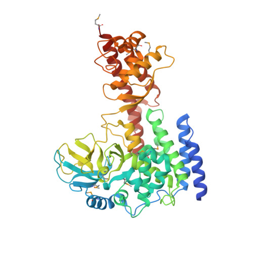 | |
UniProt & NIH Common Fund Data Resources | |||||
PHAROS: Q86TU7 GTEx: ENSG00000183576 | |||||
Entity Groups | |||||
| Sequence Clusters | 30% Identity50% Identity70% Identity90% Identity95% Identity100% Identity | ||||
| UniProt Group | Q86TU7 | ||||
Sequence AnnotationsExpand | |||||
Reference Sequence | |||||
| Ligands 5 Unique | |||||
|---|---|---|---|---|---|
| ID | Chains | Name / Formula / InChI Key | 2D Diagram | 3D Interactions | |
| SAM Download:Ideal Coordinates CCD File | B [auth A] | S-ADENOSYLMETHIONINE C15 H22 N6 O5 S MEFKEPWMEQBLKI-FCKMPRQPSA-N |  | ||
| PG4 Download:Ideal Coordinates CCD File | E [auth A] | TETRAETHYLENE GLYCOL C8 H18 O5 UWHCKJMYHZGTIT-UHFFFAOYSA-N |  | ||
| ARS Download:Ideal Coordinates CCD File | C [auth A], D [auth A] | ARSENIC As RBFQJDQYXXHULB-UHFFFAOYSA-N |  | ||
| ACT Download:Ideal Coordinates CCD File | F [auth A], G [auth A] | ACETATE ION C2 H3 O2 QTBSBXVTEAMEQO-UHFFFAOYSA-M |  | ||
| UNX Download:Ideal Coordinates CCD File | H [auth A] I [auth A] J [auth A] K [auth A] L [auth A] | UNKNOWN ATOM OR ION X |  | ||
| Modified Residues 1 Unique | |||||
|---|---|---|---|---|---|
| ID | Chains | Type | Formula | 2D Diagram | Parent |
| MSE Query on MSE | A | L-PEPTIDE LINKING | C5 H11 N O2 Se |  | MET |
| Length ( Å ) | Angle ( ˚ ) |
|---|---|
| a = 113.864 | α = 90 |
| b = 113.864 | β = 90 |
| c = 83.57 | γ = 120 |
| Software Name | Purpose |
|---|---|
| SBC-Collect | data collection |
| MOLREP | phasing |
| BUSTER | refinement |
| Coot | model building |
| HKL-2000 | data reduction |
| HKL-2000 | data scaling |


