3SKJ | pdb_00003skj

Structural And Functional Characterization of an Agonistic Anti-Human EphA2 Monoclonal Antibody


Experimental Data Snapshot

  • Method: X-RAY DIFFRACTION
  • Resolution: 2.50 Å
  • R-Value Free: 
    0.271 (Depositor), 0.273 (DCC) 
  • R-Value Work: 
    0.216 (Depositor), 0.221 (DCC) 
  • R-Value Observed: 
    0.219 (Depositor) 

wwPDB Validation 3D Report Full Report

Validation slider image for 3SKJ

This is version 1.4 of the entry. See complete history

Literature

Structural and functional characterization of an agonistic anti-human EphA2 monoclonal antibody.

Peng, L.Oganesyan, V.Damschroder, M.M.Wu, H.Dall'Acqua, W.F.

(2011) J Mol Biology 413: 390-405

  • DOI: https://doi.org/10.1016/j.jmb.2011.08.018
  • Primary Citation Related Structures: 
    3SKJ

  • PubMed Abstract: 

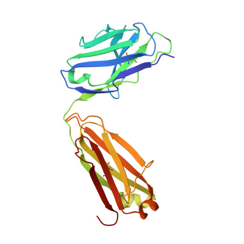
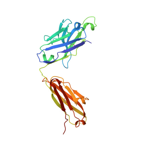
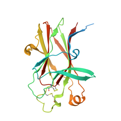
    We report here the three-dimensional structure of human ephrin type A receptor 2 (EphA2) bound to the Fab (fragment antigen binding) of an agonistic human antibody (1C1; IgG1/κ). The structure of the corresponding complex was solved at a resolution of 2.5 Å using molecular replacement and constitutes the first reported structure of a human ephrin receptor bound to an antibody. We have also defined the corresponding functional epitope using a mutagenesis-based approach. This study revealed discrete structural features that determine the fine specificity of 1C1 to EphA2. Our data also provided a molecular basis for 1C1 mechanism of action. More precisely, we propose that its agonistic, internalizing properties are the result of ligand mimicry by the third heavy-chain complementarity-determining region of 1C1. Because EphA2 is an important contributor to cancer formation and progression, these findings may have implications for designing the next generation of anti-tumor therapies.


  • Organizational Affiliation
    • Department of Antibody Discovery and Protein Engineering, MedImmune, Gaithersburg, MD 20878, USA.

Macromolecule Content 

  • Total Structure Weight: 142.94 kDa 
  • Atom Count: 8,819 
  • Modeled Residue Count: 1,144 
  • Deposited Residue Count: 1,302 
  • Unique protein chains: 3

Macromolecules

Find similar proteins by:|  3D Structure
Entity ID: 1
MoleculeChains  Sequence LengthOrganismDetailsImage
Antibody, light chainA [auth L],
C [auth M]
214Homo sapiensMutation(s): 0 
Find similar proteins by:|  3D Structure
Entity ID: 2
MoleculeChains  Sequence LengthOrganismDetailsImage
Antibody, heavy chainB [auth H],
D [auth I]
231Homo sapiensMutation(s): 0 
Find similar proteins by:|  3D Structure
Entity ID: 3
MoleculeChains  Sequence LengthOrganismDetailsImage
Ephrin type-A receptor 2
E, F
206Homo sapiensMutation(s): 0 
Gene Names: EPHA2ECK
EC: 2.7.10.1
UniProt & NIH Common Fund Data Resources
Find proteins for P29317 (Homo sapiens)
Explore P29317 
Go to UniProtKB:  P29317
PHAROS:  P29317
GTEx:  ENSG00000142627 
Entity Groups
Sequence Clusters30% Identity50% Identity70% Identity90% Identity95% Identity100% Identity
UniProt GroupP29317
Sequence Annotations
Expand
Reference Sequence

Experimental Data & Validation

Experimental Data

  • Method: X-RAY DIFFRACTION
  • Resolution: 2.50 Å
  • R-Value Free:  0.271 (Depositor), 0.273 (DCC) 
  • R-Value Work:  0.216 (Depositor), 0.221 (DCC) 
  • R-Value Observed: 0.219 (Depositor) 
Space Group: C 2 2 21
Unit Cell:
Length ( Å )Angle ( ˚ )
a = 78.94α = 90
b = 120.859β = 90
c = 286.238γ = 90
Software Package:
Software NamePurpose
ADSCdata collection
PHASESphasing
REFMACrefinement
HKL-2000data reduction
SCALEPACKdata scaling

Structure Validation

View Full Validation Report


Entry History 

Deposition Data

Revision History  (Full details and data files)

  • Version 1.0: 2011-08-31
    Type: Initial release
  • Version 1.1: 2011-09-07
    Changes: Database references
  • Version 1.2: 2012-08-29
    Changes: Database references
  • Version 1.3: 2017-05-31
    Changes: Structure summary
  • Version 1.4: 2024-11-27
    Changes: Data collection, Database references, Structure summary