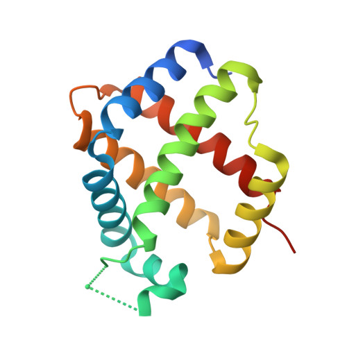
Crystal structures of Parasponia and Trema hemoglobins: differential heme coordination is linked to quaternary structure.
Kakar, S., Sturms, R., Tiffany, A., Nix, J.C., DiSpirito, A.A., Hargrove, M.S.(2011) Biochemistry 50: 4273-4280
- PubMed: 21491905
- DOI: https://doi.org/10.1021/bi2002423
- Primary Citation of Related Structures:
3QQQ, 3QQR - PubMed Abstract:
Hemoglobins from the plants Parasponia andersonii (ParaHb) and Trema tomentosa (TremaHb) are 93% identical in primary structure but differ in oxygen binding constants in accordance with their distinct physiological functions. Additionally, these proteins are dimeric, and ParaHb exhibits the unusual property of having different heme redox potentials for each subunit. To investigate how these hemoglobins could differ in function despite their shared sequence identity and to determine the cause of subunit heterogeneity in ParaHb, we have measured their crystal structures in the ferric oxidation state. Furthermore, we have made a monomeric ParaHb mutant protein (I43N) and measured its ferrous/ferric heme redox potential to test the hypothesized link between quaternary structure and heme heterogeneity in wild-type ParaHb. Our results demonstrate that TremaHb is a symmetric dimeric hemoglobin similar to other class 1 nonsymbiotic plant hemoglobins but that ParaHb has structurally distinct heme coordination in each of its two subunits that is absent in the monomeric I43N mutant protein. A mechanism for achieving structural heterogeneity in ParaHb in which the Ile(101(F4)) side chain contacts the proximal His(105(F8)) in one subunit but not the other is proposed. These results are discussed in the context of the evolution of plant oxygen transport hemoglobins, and other potential functions of plant hemoglobins.
Organizational Affiliation:
Department of Biochemistry, Biophysics, and Molecular Biology, Iowa State University, Ames, IA 50011, USA.


