3PRA | pdb_00003pra

Structural analysis of protein folding by the Methanococcus jannaschii chaperone FKBP26


Experimental Data Snapshot

  • Method: X-RAY DIFFRACTION
  • Resolution: 2.40 Å
  • R-Value Free: 
    0.291 (Depositor), 0.273 (DCC) 
  • R-Value Work: 
    0.234 (Depositor), 0.225 (DCC) 
  • R-Value Observed: 
    0.237 (Depositor) 

wwPDB Validation 3D Report Full Report

Validation slider image for 3PRA

This is version 1.3 of the entry. See complete history

Literature

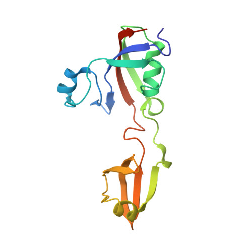
Structural Analysis of Protein Folding by the Long-Chain Archaeal Chaperone FKBP26.

Martinez-Hackert, E.Hendrickson, W.A.

(2011) J Mol Biology 407: 450-464

  • DOI: https://doi.org/10.1016/j.jmb.2011.01.027
  • Primary Citation Related Structures: 
    3PR9, 3PRA, 3PRB, 3PRD

  • PubMed Abstract: 

    In the cell, protein folding is mediated by folding catalysts and chaperones. The two functions are often linked, especially when the catalytic module forms part of a multidomain protein, as in Methanococcus jannaschii peptidyl-prolyl cis/trans isomerase FKBP26. Here, we show that FKBP26 chaperone activity requires both a 50-residue insertion in the catalytic FKBP domain, also called 'Insert-in-Flap' or IF domain, and an 80-residue C-terminal domain. We determined FKBP26 structures from four crystal forms and analyzed chaperone domains in light of their ability to mediate protein-protein interactions. FKBP26 is a crescent-shaped homodimer. We reason that folding proteins are bound inside the large crescent cleft, thus enabling their access to inward-facing peptidyl-prolyl cis/trans isomerase catalytic sites and ipsilateral chaperone domain surfaces. As these chaperone surfaces participate extensively in crystal lattice contacts, we speculate that the observed lattice contacts reflect a proclivity for protein associations and represent substrate interactions by FKBP26 chaperone domains. Finally, we find that FKBP26 is an exceptionally flexible molecule, suggesting a mechanism for nonspecific substrate recognition.


  • Organizational Affiliation
    • Department of Biochemistry and Molecular Biophysics, Columbia University, New York, NY 10032, USA.

Macromolecule Content 

  • Total Structure Weight: 35.02 kDa 
  • Atom Count: 2,480 
  • Modeled Residue Count: 300 
  • Deposited Residue Count: 314 
  • Unique protein chains: 1

Macromolecules

Find similar proteins by:|  3D Structure
Entity ID: 1
MoleculeChains  Sequence LengthOrganismDetailsImage
FKBP-type peptidyl-prolyl cis-trans isomerase
A, B
157Methanocaldococcus jannaschiiMutation(s): 0 
Gene Names: MJ0825
EC: 5.2.1.8
UniProt
Find proteins for Q58235 (Methanocaldococcus jannaschii (strain ATCC 43067 / DSM 2661 / JAL-1 / JCM 10045 / NBRC 100440))
Explore Q58235 
Go to UniProtKB:  Q58235
Entity Groups
Sequence Clusters30% Identity50% Identity70% Identity90% Identity95% Identity100% Identity
UniProt GroupQ58235
Sequence Annotations
Expand
Reference Sequence

Experimental Data & Validation

Experimental Data

  • Method: X-RAY DIFFRACTION
  • Resolution: 2.40 Å
  • R-Value Free:  0.291 (Depositor), 0.273 (DCC) 
  • R-Value Work:  0.234 (Depositor), 0.225 (DCC) 
  • R-Value Observed: 0.237 (Depositor) 
Space Group: P 1 21 1
Unit Cell:
Length ( Å )Angle ( ˚ )
a = 45.984α = 90
b = 67.886β = 95.29
c = 54.466γ = 90
Software Package:
Software NamePurpose
REFMACrefinement
SCALEPACKdata scaling
CNSrefinement
DENZOdata reduction
PDB_EXTRACTdata extraction
CNSphasing

Structure Validation

View Full Validation Report


Entry History 

Deposition Data

Revision History  (Full details and data files)

  • Version 1.0: 2011-01-19
    Type: Initial release
  • Version 1.1: 2011-07-13
    Changes: Version format compliance
  • Version 1.2: 2019-07-17
    Changes: Data collection, Refinement description
  • Version 1.3: 2024-02-21
    Changes: Data collection, Database references