3PLT | pdb_00003plt

Crystal structure of Lsp1 from Saccharomyces cerevisiae


Experimental Data Snapshot

  • Method: X-RAY DIFFRACTION
  • Resolution: 2.90 Å
  • R-Value Free: 
    0.296 (Depositor), 0.284 (DCC) 
  • R-Value Work: 
    0.236 (Depositor), 0.232 (DCC) 
  • R-Value Observed: 
    0.239 (Depositor) 

wwPDB Validation 3D Report Full Report

Validation slider image for 3PLT

This is version 1.3 of the entry. See complete history

Literature

Eisosome-driven plasma membrane organization is mediated by BAR domains.

Ziolkowska, N.E.Karotki, L.Rehman, M.Huiskonen, J.T.Walther, T.C.

(2011) Nat Struct Mol Biol 18: 854-856

  • DOI: https://doi.org/10.1038/nsmb.2080
  • Primary Citation Related Structures: 
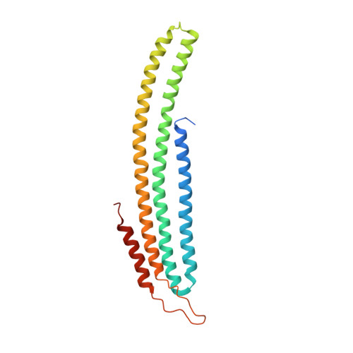
    3PLT

  • PubMed Abstract: 

    Plasma membranes are organized into domains of different protein and lipid composition. Eisosomes are key complexes for yeast plasma membrane organization, containing primarily Pil1 and Lsp1. Here we show that both proteins consist mostly of a banana-shaped BAR domain common to membrane sculpting proteins, most similar to the ones of amphiphysin, arfaptin 2 and endophilin 2. Our data reveal a previously unrecognized family of BAR-domain proteins involved in plasma membrane organization.


  • Organizational Affiliation
    • Max Planck Institute of Biochemistry, Organelle Architecture and Dynamics, Martinsried, Germany.

Macromolecule Content 

  • Total Structure Weight: 78.24 kDa 
  • Atom Count: 5,037 
  • Modeled Residue Count: 634 
  • Deposited Residue Count: 702 
  • Unique protein chains: 1

Macromolecules

Find similar proteins by:|  3D Structure
Entity ID: 1
MoleculeChains  Sequence LengthOrganismDetailsImage
Sphingolipid long chain base-responsive protein LSP1
A, B, C
234Saccharomyces cerevisiaeMutation(s): 0 
Gene Names: LSP1YPL004C
UniProt
Find proteins for Q12230 (Saccharomyces cerevisiae (strain ATCC 204508 / S288c))
Explore Q12230 
Go to UniProtKB:  Q12230
Entity Groups
Sequence Clusters30% Identity50% Identity70% Identity90% Identity95% Identity100% Identity
UniProt GroupQ12230
Sequence Annotations
Expand
Reference Sequence

Experimental Data & Validation

Experimental Data

  • Method: X-RAY DIFFRACTION
  • Resolution: 2.90 Å
  • R-Value Free:  0.296 (Depositor), 0.284 (DCC) 
  • R-Value Work:  0.236 (Depositor), 0.232 (DCC) 
  • R-Value Observed: 0.239 (Depositor) 
Space Group: C 1 2 1
Unit Cell:
Length ( Å )Angle ( ˚ )
a = 273.16α = 90
b = 38.753β = 99.349
c = 75.509γ = 90
Software Package:
Software NamePurpose
HKL-3000phasing
MOLREPphasing
REFMACrefinement
HKL-2000data reduction
HKL-3000data scaling

Structure Validation

View Full Validation Report


Entry History 

Deposition Data

Revision History  (Full details and data files)

  • Version 1.0: 2011-06-22
    Type: Initial release
  • Version 1.1: 2011-07-13
    Changes: Version format compliance
  • Version 1.2: 2011-07-20
    Changes: Database references
  • Version 1.3: 2024-02-21
    Changes: Data collection, Database references, Refinement description