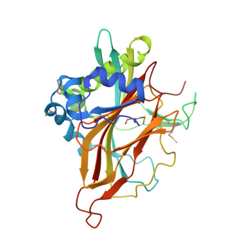
3PL0 | pdb_00003pl0

Crystal structure of a bsmA homolog (Mpe_A2762) from Methylobium petroleophilum PM1 at 1.91 A resolution


Experimental Data Snapshot

  • Method: X-RAY DIFFRACTION
  • Resolution: 1.91 Å
  • R-Value Free: 
    0.211 (Depositor), 0.207 (DCC) 
  • R-Value Work: 
    0.169 (Depositor), 0.172 (DCC) 
  • R-Value Observed: 
    0.172 (Depositor) 

wwPDB Validation 3D Report Full Report

Validation slider image for 3PL0

This is version 1.5 of the entry. See complete history. 

Literature

Crystal structure of a member of a novel family of dioxygenases (PF10014) reveals a conserved cupin fold and active site.

Xu, Q., Grant, J., Chiu, H.J., Farr, C.L., Jaroszewski, L., Knuth, M.W., Miller, M.D., Lesley, S.A., Godzik, A., Elsliger, M.A., Deacon, A.M., Wilson, I.A.

(2014) Proteins 82: 164-170

  • DOI: https://doi.org/10.1002/prot.24362
  • Primary Citation Related Structures: 
    3PL0

  • PubMed Abstract: 

    PF10014 is a novel family of 2-oxyglutarate-Fe(2+) -dependent dioxygenases that are involved in biosynthesis of antibiotics and regulation of biofilm formation, likely by catalyzing hydroxylation of free amino acids or other related ligands. The crystal structure of a PF10014 member from Methylibium petroleiphilum at 1.9 Å resolution shows strong structural similarity to cupin dioxygenases in overall fold and active site, despite very remote homology. However, one of the β-strands of the cupin catalytic core is replaced by a loop that displays conformational isomerism that likely regulates the active site.


  • Organizational Affiliation: 
    • Joint Center for Structural Genomics, La Jolla, California (http://www.jcsg.org); Stanford Synchrotron Radiation Lightsource, SLAC National Accelerator Laboratory, Menlo Park, California, 94025.

Macromolecule Content 

  • Total Structure Weight: 57.41 kDa 
  • Atom Count: 4,516 
  • Modeled Residue Count: 506 
  • Deposited Residue Count: 508 
  • Unique protein chains: 1

Macromolecules

Find similar proteins by:|  3D Structure
Entity ID: 1
MoleculeChains  Sequence LengthOrganismDetailsImage
Uncharacterized protein
A, B
254Methylibium petroleiphilum PM1Mutation(s): 0 
Gene Names: Mpe_A2762
UniProt
Find proteins for A2SJH7 (Methylibium petroleiphilum (strain ATCC BAA-1232 / LMG 22953 / PM1))
Explore A2SJH7 
Go to UniProtKB:  A2SJH7
Entity Groups
Sequence Clusters30% Identity50% Identity70% Identity90% Identity95% Identity100% Identity
UniProt GroupA2SJH7
Sequence Annotations
Expand
Reference Sequence

Small Molecules

Modified Residues  1 Unique
IDChains TypeFormula2D DiagramParent
MSE
Query on MSE
A, B
L-PEPTIDE LINKINGC5 H11 N O2 SeMET

Experimental Data & Validation

Experimental Data

  • Method: X-RAY DIFFRACTION
  • Resolution: 1.91 Å
  • R-Value Free:  0.211 (Depositor), 0.207 (DCC) 
  • R-Value Work:  0.169 (Depositor), 0.172 (DCC) 
  • R-Value Observed: 0.172 (Depositor) 
Space Group: P 1 21 1
Unit Cell:
Length ( Å )Angle ( ˚ )
a = 43.508α = 90
b = 63.203β = 97.8
c = 104.504γ = 90
Software Package:
Software NamePurpose
SHELXphasing
REFMACrefinement
XSCALEdata scaling
PDB_EXTRACTdata extraction
XDSdata reduction
SHELXDphasing
autoSHARPphasing

Structure Validation

View Full Validation Report


Entry History 

Deposition Data

Revision History  (Full details and data files)

  • Version 1.0: 2010-12-08
    Type: Initial release
  • Version 1.1: 2011-07-13
    Changes: Version format compliance
  • Version 1.2: 2011-07-20
    Changes: Structure summary
  • Version 1.3: 2014-09-24
    Changes: Database references
  • Version 1.4: 2023-02-01
    Changes: Database references, Derived calculations
  • Version 1.5: 2024-11-20
    Changes: Data collection, Refinement description, Structure summary