3P3N | pdb_00003p3n

Factor inhibiting HIF-1 Alpha in complex with Notch 1 fragment mouse notch (1930-1949) peptide


Experimental Data Snapshot

  • Method: X-RAY DIFFRACTION
  • Resolution: 2.40 Å
  • R-Value Free: 
    0.244 (Depositor), 0.221 (DCC) 
  • R-Value Work: 
    0.204 (Depositor), 0.204 (DCC) 
  • R-Value Observed: 
    0.204 (Depositor) 

Starting Model: experimental
View more details

wwPDB Validation 3D Report Full Report

Validation slider image for 3P3N

This is version 1.2 of the entry. See complete history

Literature

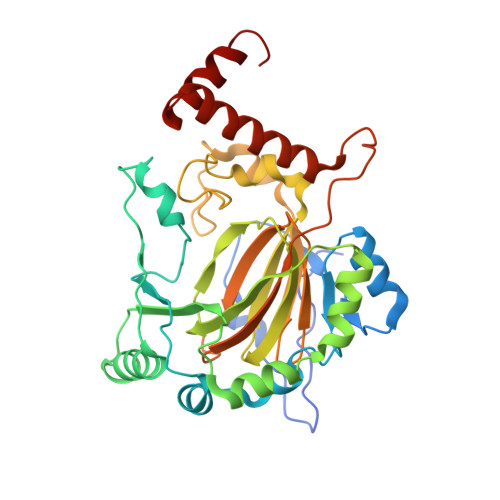
Asparaginyl hydroxylation of the Notch ankyrin repeat domain by factor inhibiting hypoxia-inducible factor

Coleman, M.L.Mcdonough, M.A.Hewitson, K.S.Coles, C.Mecinovic, J.Edelmann, M.Cook, K.M.Cockman, M.E.Lancaster, D.E.Kessler, B.M.Oldham, N.J.Ratcliffe, P.J.Schofield, C.J.

(2007) J Biological Chem 282: 24027-24038

  • DOI: https://doi.org/10.1074/jbc.M704102200
  • Primary Citation Related Structures: 
    2QC9, 3P3N, 3P3P

  • PubMed Abstract: 

    The stability and activity of hypoxia-inducible factor (HIF) are regulated by the post-translational hydroxylation of specific prolyl and asparaginyl residues. We show that the HIF asparaginyl hydroxylase, factor inhibiting HIF (FIH), also catalyzes hydroxylation of highly conserved asparaginyl residues within ankyrin repeat (AR) domains (ARDs) of endogenous Notch receptors. AR hydroxylation decreases the extent of ARD binding to FIH while not affecting signaling through the canonical Notch pathway. ARD proteins were found to efficiently compete with HIF for FIH-dependent hydroxylation. Crystallographic analyses of the hydroxylated Notch ARD (2.35A) and of Notch peptides bound to FIH (2.4-2.6A) reveal the stereochemistry of hydroxylation on the AR and imply that significant conformational changes are required in the ARD fold in order to enable hydroxylation at the FIH active site. We propose that ARD proteins function as natural inhibitors of FIH and that the hydroxylation status of these proteins provides another oxygen-dependent interface that modulates HIF signaling.


  • Organizational Affiliation
    • Henry Wellcome Building for Molecular Physiology, University of Oxford, Oxford, OX3 7BN, United Kingdom.

Macromolecule Content 

  • Total Structure Weight: 43.25 kDa 
  • Atom Count: 2,990 
  • Modeled Residue Count: 351 
  • Deposited Residue Count: 369 
  • Unique protein chains: 2

Macromolecules

Find similar proteins by:|  3D Structure
Entity ID: 1
MoleculeChains  Sequence LengthOrganismDetailsImage
Hypoxia-inducible factor 1-alpha inhibitor349Homo sapiensMutation(s): 0 
Gene Names: FIH1HIF1AN
EC: 1.14.11.16 (PDB Primary Data), 1.14.11.30 (UniProt)
UniProt & NIH Common Fund Data Resources
Find proteins for Q9NWT6 (Homo sapiens)
Explore Q9NWT6 
Go to UniProtKB:  Q9NWT6
PHAROS:  Q9NWT6
GTEx:  ENSG00000166135 
Entity Groups
Sequence Clusters30% Identity50% Identity70% Identity90% Identity95% Identity100% Identity
UniProt GroupQ9NWT6
Sequence Annotations
Expand
Reference Sequence
Find similar proteins by:  Sequence   |   3D Structure  
Entity ID: 2
MoleculeChains  Sequence LengthOrganismDetailsImage
Notch 1 protein20Mus musculusMutation(s): 0 
UniProt & NIH Common Fund Data Resources
Find proteins for Q01705 (Mus musculus)
Explore Q01705 
Go to UniProtKB:  Q01705
IMPC:  MGI:97363
Entity Groups
Sequence Clusters30% Identity50% Identity70% Identity90% Identity95% Identity100% Identity
UniProt GroupQ01705
Sequence Annotations
Expand
Reference Sequence

Small Molecules

Ligands 4 Unique
IDChains Name / Formula / InChI Key2D Diagram3D Interactions
AKG

Query on AKG


Download:Ideal Coordinates CCD File
F [auth A]2-OXOGLUTARIC ACID
C5 H6 O5
KPGXRSRHYNQIFN-UHFFFAOYSA-N
SO4

Query on SO4


Download:Ideal Coordinates CCD File
D [auth A],
E [auth A]
SULFATE ION
O4 S
QAOWNCQODCNURD-UHFFFAOYSA-L
GOL

Query on GOL


Download:Ideal Coordinates CCD File
G [auth A],
H [auth A],
I [auth A],
J [auth A]
GLYCEROL
C3 H8 O3
PEDCQBHIVMGVHV-UHFFFAOYSA-N
FE2

Query on FE2


Download:Ideal Coordinates CCD File
C [auth A]FE (II) ION
Fe
CWYNVVGOOAEACU-UHFFFAOYSA-N

Experimental Data & Validation

Experimental Data

  • Method: X-RAY DIFFRACTION
  • Resolution: 2.40 Å
  • R-Value Free:  0.244 (Depositor), 0.221 (DCC) 
  • R-Value Work:  0.204 (Depositor), 0.204 (DCC) 
  • R-Value Observed: 0.204 (Depositor) 
Space Group: P 41 21 2
Unit Cell:
Length ( Å )Angle ( ˚ )
a = 86.188α = 90
b = 86.188β = 90
c = 147.943γ = 90
Software Package:
Software NamePurpose
ADSCdata collection
CNSrefinement
HKL-2000data reduction
HKL-2000data scaling
CNSphasing

Structure Validation

View Full Validation Report


Entry History 

Deposition Data

Revision History  (Full details and data files)

  • Version 1.0: 2010-11-24
    Type: Initial release
  • Version 1.1: 2011-07-13
    Changes: Version format compliance
  • Version 1.2: 2023-09-06
    Changes: Data collection, Database references, Derived calculations, Refinement description