3OB4 | pdb_00003ob4

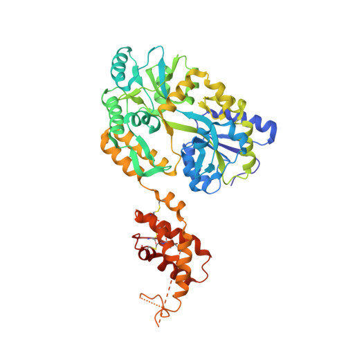
MBP-fusion protein of the major peanut allergen Ara h 2


Experimental Data Snapshot

  • Method: X-RAY DIFFRACTION
  • Resolution: 2.71 Å
  • R-Value Free: 
    0.256 (Depositor), 0.251 (DCC) 
  • R-Value Work: 
    0.183 (Depositor), 0.177 (DCC) 
  • R-Value Observed: 
    0.187 (Depositor) 

Starting Model: experimental
View more details

wwPDB Validation 3D Report Full Report

Validation slider image for 3OB4

This is version 2.3 of the entry. See complete history. 

Literature

Ara h 2: crystal structure and IgE binding distinguish two subpopulations of peanut allergic patients by epitope diversity.

Mueller, G.A., Gosavi, R.A., Pomes, A., Wunschmann, S., Moon, A.F., London, R.E., Pedersen, L.C.

(2011) Allergy 66: 878-885

  • DOI: https://doi.org/10.1111/j.1398-9995.2010.02532.x
  • Primary Citation Related Structures: 
    3OB4

  • PubMed Abstract: 

    Peanut allergy affects 1% of the population and causes the most fatal food-related anaphylactic reactions. The protein Ara h 2 is the most potent peanut allergen recognized by 80-90% of peanut allergic patients. The crystal structure of the major peanut allergen Ara h 2 was determined for the first time at 2.7 Å resolution using a customized maltose-binding protein (MBP)-fusion system. IgE antibody binding to the MBP fusion construct vs the natural allergen was compared by ELISA using sera from peanut allergic patients. The structure of Ara h 2 is a five-helix bundle held together by four disulfide bonds and related to the prolamin protein superfamily. The fold is most similar to other amylase and trypsin inhibitors. The MBP--Ara h 2 fusion construct was positively recognized by IgE from 76% of allergic patients (25/33). Two populations of patients could be identified. Subpopulation 1 (n = 14) showed an excellent correlation of IgE antibody binding to natural vs recombinant Ara h 2. Subpopulation 2 (n = 15) showed significantly reduced IgE binding to the MBP fusion protein. Interestingly, about 20% of the IgE binding in subpopulation 2 could be recovered by increasing the distance between MBP and Ara h 2 in a second construct. The reduced IgE binding to the MBP--Ara h 2 of subpopulation 2 indicates that the MBP molecule protects an immunodominant epitope region near the first helix of Ara h 2. Residues involved in the epitope(s) are suggested by the crystal structure. The MBP--Ara h 2 fusion constructs will be useful to further elucidate the relevance of certain epitopes to peanut allergy.


  • Organizational Affiliation: 
    • Laboratory of Structural Biology, National Institute of Environmental Health Sciences, Research Triangle Park, NC, USA. mueller3@niehs.nih.gov

Macromolecule Content 

  • Total Structure Weight: 56.63 kDa 
  • Atom Count: 3,760 
  • Modeled Residue Count: 474 
  • Deposited Residue Count: 500 
  • Unique protein chains: 1

Macromolecules

Find similar proteins by:|  3D Structure
Entity ID: 1
MoleculeChains  Sequence LengthOrganismDetailsImage
Maltose ABC transporter periplasmic protein,Arah 2500Escherichia coli, Arachis hypogaea
This entity is chimeric
Mutation(s): 7 
Gene Names: HMPREF9530_03068, Ara h 2
UniProt
Find proteins for A5Z1Q8 (Arachis duranensis)
Explore A5Z1Q8 
Go to UniProtKB:  A5Z1Q8
Find proteins for P0AEX9 (Escherichia coli (strain K12))
Explore P0AEX9 
Go to UniProtKB:  P0AEX9
Entity Groups
Sequence Clusters30% Identity50% Identity70% Identity90% Identity95% Identity100% Identity
UniProt GroupsP0AEX9A5Z1Q8
Sequence Annotations
Expand
Reference Sequence

Oligosaccharides

Help  
Entity ID: 2
MoleculeChains Length2D Diagram GlycosylationD Interactions
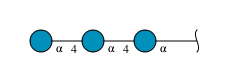
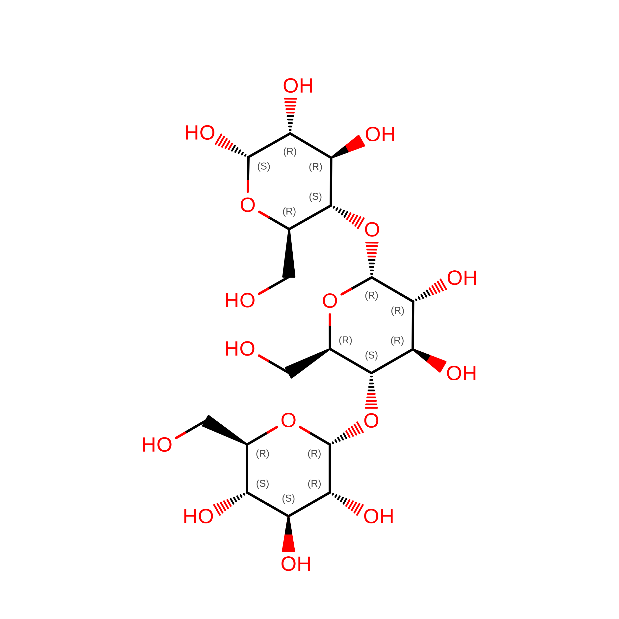
alpha-D-glucopyranose-(1-4)-alpha-D-glucopyranose-(1-4)-alpha-D-glucopyranose
B
3N/A
Glycosylation Resources
GlyTouCan: G96370VA
GlyCosmos: G96370VA
GlyGen: G96370VA

Small Molecules

Ligands 2 Unique
IDChains Name / Formula / InChI Key2D Diagram3D Interactions
SO4

Query on SO4


Download:Ideal Coordinates CCD File
C [auth A],
D [auth A],
E [auth A]
SULFATE ION
O4 S
QAOWNCQODCNURD-UHFFFAOYSA-L
CL

Query on CL


Download:Ideal Coordinates CCD File
F [auth A],
G [auth A],
H [auth A],
I [auth A],
J [auth A]
CHLORIDE ION
Cl
VEXZGXHMUGYJMC-UHFFFAOYSA-M

Biologically Interesting Molecules (External Reference) 

1 Unique

Experimental Data & Validation

Experimental Data

  • Method: X-RAY DIFFRACTION
  • Resolution: 2.71 Å
  • R-Value Free:  0.256 (Depositor), 0.251 (DCC) 
  • R-Value Work:  0.183 (Depositor), 0.177 (DCC) 
  • R-Value Observed: 0.187 (Depositor) 
Space Group: C 1 2 1
Unit Cell:
Length ( Å )Angle ( ˚ )
a = 68.847α = 90
b = 87.381β = 103.93
c = 113.137γ = 90
Software Package:
Software NamePurpose
MAR345data collection
MOLREPphasing
PHENIXrefinement
HKL-2000data reduction
HKL-2000data scaling

Structure Validation

View Full Validation Report


Entry History 

Deposition Data

Revision History  (Full details and data files)

  • Version 1.0: 2011-02-02
    Type: Initial release
  • Version 1.1: 2011-07-13
    Changes: Version format compliance
  • Version 1.2: 2017-08-09
    Changes: Refinement description, Source and taxonomy
  • Version 1.3: 2017-11-08
    Changes: Refinement description
  • Version 1.4: 2020-02-26
    Changes: Database references
  • Version 2.0: 2020-07-29
    Type: Remediation
    Reason: Carbohydrate remediation
    Changes: Atomic model, Data collection, Derived calculations, Non-polymer description, Structure summary
  • Version 2.1: 2021-02-17
    Changes: Database references, Source and taxonomy, Structure summary
  • Version 2.2: 2023-09-06
    Changes: Data collection, Database references, Refinement description
  • Version 2.3: 2024-10-09
    Changes: Structure summary