3O7R | pdb_00003o7r

Crystal structure of Ru(p-cymene)/apo-H49AFr


Experimental Data Snapshot

  • Method: X-RAY DIFFRACTION
  • Resolution: 1.90 Å
  • R-Value Free: 
    0.237 (Depositor), 0.236 (DCC) 
  • R-Value Work: 
    0.193 (Depositor) 
  • R-Value Observed: 
    0.196 (Depositor) 

wwPDB Validation 3D Report Full Report

Validation slider image for 3O7R

This is version 1.2 of the entry. See complete history

Literature

Incorporation of organometallic Ru complexes into apo-ferritin cage.

Takezawa, Y.Bockmann, P.Sugi, N.Wang, Z.Abe, S.Murakami, T.Hikage, T.Erker, G.Watanabe, Y.Kitagawa, S.Ueno, T.

(2011) J Chem Soc ,dalton Trans 40: 2190-2195

  • DOI: https://doi.org/10.1039/c0dt00955e
  • Primary Citation Related Structures: 
    3O7R, 3O7S

  • PubMed Abstract: 

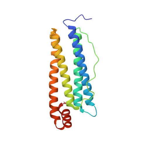
    Spherical protein cages such as an iron storage protein, ferritin, have great potential as nanometer-scale capsules to assemble and store metal ions and complexes. We report herein the synthesis of a composite of an apo-ferritin cage and Ru(p-cymene) complexes. Ru complexes were efficiently incorporated into the ferritin cavity without degradation of its cage structure. X-Ray crystallography revealed that the Ru complexes were immobilized on the interior surface of the cage mainly by the coordination of histidine residues.


  • Organizational Affiliation
    • Research Center for Materials Science, Nagoya University, Furo-cho, Nagoya, Japan.

Macromolecule Content 

  • Total Structure Weight: 20.89 kDa 
  • Atom Count: 1,640 
  • Modeled Residue Count: 173 
  • Deposited Residue Count: 174 
  • Unique protein chains: 1

Macromolecules

Find similar proteins by:|  3D Structure
Entity ID: 1
MoleculeChains  Sequence LengthOrganismDetailsImage
Ferritin light chain174Equus caballusMutation(s): 2 
Gene Names: FTL
UniProt
Find proteins for P02791 (Equus caballus)
Explore P02791 
Go to UniProtKB:  P02791
Entity Groups
Sequence Clusters30% Identity50% Identity70% Identity90% Identity95% Identity100% Identity
UniProt GroupP02791
Sequence Annotations
Expand
Reference Sequence

Small Molecules

Ligands 4 Unique
IDChains Name / Formula / InChI Key2D Diagram3D Interactions
CD

Query on CD


Download:Ideal Coordinates CCD File
B [auth A],
C [auth A],
D [auth A]
CADMIUM ION
Cd
WLZRMCYVCSSEQC-UHFFFAOYSA-N
RU

Query on RU


Download:Ideal Coordinates CCD File
E [auth A],
F [auth A]
RUTHENIUM ION
Ru
BPEVHDGLPIIAGH-UHFFFAOYSA-N
SO4

Query on SO4


Download:Ideal Coordinates CCD File
G [auth A],
H [auth A]
SULFATE ION
O4 S
QAOWNCQODCNURD-UHFFFAOYSA-L
EDO

Query on EDO


Download:Ideal Coordinates CCD File
I [auth A]
J [auth A]
K [auth A]
L [auth A]
M [auth A]
I [auth A],
J [auth A],
K [auth A],
L [auth A],
M [auth A],
N [auth A]
1,2-ETHANEDIOL
C2 H6 O2
LYCAIKOWRPUZTN-UHFFFAOYSA-N

Experimental Data & Validation

Experimental Data

  • Method: X-RAY DIFFRACTION
  • Resolution: 1.90 Å
  • R-Value Free:  0.237 (Depositor), 0.236 (DCC) 
  • R-Value Work:  0.193 (Depositor) 
  • R-Value Observed: 0.196 (Depositor) 
Space Group: F 4 3 2
Unit Cell:
Length ( Å )Angle ( ˚ )
a = 181.703α = 90
b = 181.703β = 90
c = 181.703γ = 90
Software Package:
Software NamePurpose
REFMACrefinement
PDB_EXTRACTdata extraction
HKL-2000data collection
HKL-2000data reduction
HKL-2000data scaling

Structure Validation

View Full Validation Report


Entry History 

Deposition Data

Revision History  (Full details and data files)

  • Version 1.0: 2011-04-20
    Type: Initial release
  • Version 1.1: 2011-07-13
    Changes: Version format compliance
  • Version 1.2: 2024-03-20
    Changes: Data collection, Database references, Derived calculations