3O6N | pdb_00003o6n

Crystal Structure of APL1 leucine-rich repeat domain


Experimental Data Snapshot

  • Method: X-RAY DIFFRACTION
  • Resolution: 1.85 Å
  • R-Value Free: 
    0.200 (Depositor), 0.207 (DCC) 
  • R-Value Work: 
    0.169 (Depositor), 0.179 (DCC) 
  • R-Value Observed: 
    0.171 (Depositor) 

wwPDB Validation 3D Report Full Report

Validation slider image for 3O6N

Ligand Structure Quality Assessment 


This is version 2.1 of the entry. See complete history

Literature

A heterodimeric complex of the LRR proteins LRIM1 and APL1C regulates complement-like immunity in Anopheles gambiae.

Baxter, R.H.Steinert, S.Chelliah, Y.Volohonsky, G.Levashina, E.A.Deisenhofer, J.

(2010) Proc Natl Acad Sci U S A 107: 16817-16822

  • DOI: https://doi.org/10.1073/pnas.1010575107
  • Primary Citation Related Structures: 
    3O53, 3O6N, 3OJA

  • PubMed Abstract: 

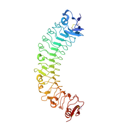
    The leucine-rich repeat (LRR) proteins LRIM1 and APL1C control the function of the complement-like protein TEP1 in Anopheles mosquitoes. The molecular structure of LRIM1 and APL1C and the basis of their interaction with TEP1 represent a new type of innate immune complex. The LRIM1/APL1C complex specifically binds and solubilizes a cleaved form of TEP1 without an intact thioester bond. The LRIM1 and APL1C LRR domains have a large radius of curvature, glycosylated concave face, and a novel C-terminal capping motif. The LRIM1/APL1C complex is a heterodimer with a single intermolecular disulfide bond. The structure of the LRIM1/APL1C heterodimer reveals an interface between the two LRR domains and an extensive C-terminal coiled-coil domain. We propose that a cleaved form of TEP1 may act as a convertase for activation of other TEP1 molecules and that the LRIM1/APL1C heterodimer regulates formation of this TEP1 convertase.


  • Organizational Affiliation
    • Howard Hughes Medical Institute and Department of Biochemistry, University of Texas Southwestern Medical Center, 6001 Forest Park Road, Dallas, TX 75390-9050, USA.

Macromolecule Content 

  • Total Structure Weight: 46 kDa 
  • Atom Count: 3,597 
  • Modeled Residue Count: 378 
  • Deposited Residue Count: 390 
  • Unique protein chains: 1

Macromolecules

Find similar proteins by:|  3D Structure
Entity ID: 1
MoleculeChains  Sequence LengthOrganismDetailsImage
APL1390Anopheles gambiaeMutation(s): 0 
Gene Names: APL1

Oligosaccharides

Help  
Entity ID: 2
MoleculeChains Length2D Diagram GlycosylationD Interactions
2-acetamido-2-deoxy-beta-D-glucopyranose-(1-4)-2-acetamido-2-deoxy-beta-D-glucopyranose
B
2N-Glycosylation
Glycosylation Resources
GlyTouCan: G42666HT
GlyCosmos: G42666HT
GlyGen: G42666HT

Experimental Data & Validation

Experimental Data

  • Method: X-RAY DIFFRACTION
  • Resolution: 1.85 Å
  • R-Value Free:  0.200 (Depositor), 0.207 (DCC) 
  • R-Value Work:  0.169 (Depositor), 0.179 (DCC) 
  • R-Value Observed: 0.171 (Depositor) 
Space Group: P 21 21 21
Unit Cell:
Length ( Å )Angle ( ˚ )
a = 38.77α = 90
b = 70.35β = 90
c = 161.76γ = 90
Software Package:
Software NamePurpose
XSCALEdata processing
PHENIXrefinement
PDB_EXTRACTdata extraction
XSCALEdata scaling

Structure Validation

View Full Validation Report


Ligand Structure Quality Assessment 


Entry History 

Deposition Data

Revision History  (Full details and data files)

  • Version 1.0: 2010-09-22
    Type: Initial release
  • Version 1.1: 2011-07-13
    Changes: Version format compliance
  • Version 2.0: 2020-07-29
    Type: Remediation
    Reason: Carbohydrate remediation
    Changes: Atomic model, Data collection, Database references, Derived calculations, Structure summary
  • Version 2.1: 2024-10-16
    Changes: Data collection, Database references, Structure summary