3NJ6 | pdb_00003nj6

0.95 A resolution X-ray structure of (GGCAGCAGCC)2


Experimental Data Snapshot

  • Method: X-RAY DIFFRACTION
  • Resolution: 0.95 Å
  • R-Value Work: 
    0.106 (Depositor), 0.102 (DCC) 
  • R-Value Observed: 
    0.106 (Depositor) 

Starting Model: in silico
View more details

wwPDB Validation 3D Report Full Report

Validation slider image for 3NJ6

This is version 1.4 of the entry. See complete history

Literature

Atomic resolution structure of CAG RNA repeats: structural insights and implications for the trinucleotide repeat expansion diseases.

Kiliszek, A.Kierzek, R.Krzyzosiak, W.J.Rypniewski, W.

(2010) Nucleic Acids Res 38: 8370-8376

  • DOI: https://doi.org/10.1093/nar/gkq700
  • Primary Citation Related Structures: 
    3NJ6, 3NJ7

  • PubMed Abstract: 

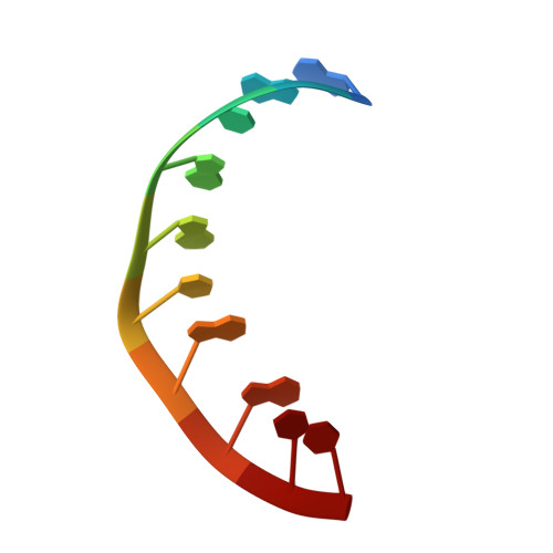
    CAG repeats occur predominantly in the coding regions of human genes, which suggests their functional importance. In some genes, these sequences can undergo pathogenic expansions leading to neurodegenerative polyglutamine (poly-Q) diseases. The mutant transcripts containing expanded CAG repeats possibly contribute to pathogenesis in addition to the well-known pathogenic effects of mutant proteins. We have analysed two crystal forms of RNA duplexes containing CAG repeats: (GGCAGCAGCC)(2). One of the structures has been determined at atomic resolution (0.95 Å) and the other at 1.9 Å. The duplexes include non-canonical A-A pairs that fit remarkably well within a regular A-helix. All the adenosines are in the anti-conformation and the only interaction within each A-A pair is a single C2-H2···N1 hydrogen bond. Both adenosines in each A-A pair are shifted towards the major groove, although to different extents; the A which is the H-bond donor stands out more (the 'thumbs-up' conformation). The main effect on the helix conformation is a local unwinding. The CAG repeats and the previously examined CUG structures share a similar pattern of electrostatic charge distribution in the minor groove, which could explain their affinity for the pathogenesis-related MBNL1 protein.


  • Organizational Affiliation
    • Institute of Bioorganic Chemistry, Polish Academy of Sciences, Noskowskiego 12/14, 61-704 Poznan, Poland.

Macromolecule Content 

  • Total Structure Weight: 3.31 kDa 
  • Atom Count: 305 
  • Modeled Residue Count: 10 
  • Deposited Residue Count: 10 
  • Unique nucleic acid chains: 1

Macromolecules

Find similar nucleic acids by:  Sequence
Entity ID: 1
MoleculeChains LengthOrganismImage
5'-R(*GP*GP*CP*AP*GP*CP*AP*GP*CP*C)-3'10N/A
Sequence Annotations
Expand
Reference Sequence

Small Molecules

Ligands 1 Unique
IDChains Name / Formula / InChI Key2D Diagram3D Interactions
SO4

Query on SO4


Download:Ideal Coordinates CCD File
B [auth A]SULFATE ION
O4 S
QAOWNCQODCNURD-UHFFFAOYSA-L

Experimental Data & Validation

Experimental Data

  • Method: X-RAY DIFFRACTION
  • Resolution: 0.95 Å
  • R-Value Work:  0.106 (Depositor), 0.102 (DCC) 
  • R-Value Observed: 0.106 (Depositor) 
Space Group: H 3 2
Unit Cell:
Length ( Å )Angle ( ˚ )
a = 46.427α = 90
b = 46.427β = 90
c = 82.678γ = 120
Software Package:
Software NamePurpose
DENZOdata reduction
SCALEPACKdata scaling
PHASERphasing
PHENIXrefinement
PDB_EXTRACTdata extraction
HKL-2000data reduction
HKL-2000data scaling

Structure Validation

View Full Validation Report


Entry History 

Deposition Data

Revision History  (Full details and data files)

  • Version 1.0: 2010-08-25
    Type: Initial release
  • Version 1.1: 2011-07-13
    Changes: Version format compliance
  • Version 1.2: 2017-11-08
    Changes: Refinement description
  • Version 1.3: 2024-02-21
    Changes: Data collection, Database references, Derived calculations
  • Version 1.4: 2024-04-03
    Changes: Refinement description