3NE9 | pdb_00003ne9

Chronobacterium ammoiniagenes apo-ACPS structure


Experimental Data Snapshot

  • Method: X-RAY DIFFRACTION
  • Resolution: 2.50 Å
  • R-Value Free: 
    0.268 (Depositor) 
  • R-Value Work: 
    0.205 (Depositor) 
  • R-Value Observed: 
    0.208 (Depositor) 

wwPDB Validation 3D Report Full Report

Validation slider image for 3NE9

This is version 1.4 of the entry. See complete history

Literature

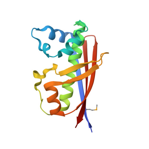
Mycobacterium tuberculosis acyl carrier protein synthase adopts two different pH-dependent structural conformations.

Gokulan, K.Aggarwal, A.Shipman, L.Besra, G.S.Sacchettini, J.C.

(2011) Acta Crystallogr D Biol Crystallogr 67: 657-669

  • DOI: https://doi.org/10.1107/S0907444911020221
  • Primary Citation Related Structures: 
    3NE1, 3NE3, 3NE9, 3NFD

  • PubMed Abstract: 

    The crystal structures of acyl carrier protein synthase (AcpS) from Mycobacterium tuberculosis (Mtb) and Corynebacterium ammoniagenes determined at pH 5.3 and pH 6.5, respectively, are reported. Comparison of the Mtb apo-AcpS structure with the recently reported structure of the Mtb AcpS-ADP complex revealed that AcpS adopts two different conformations: the orthorhombic and trigonal space-group structures show structural differences in the α2 helix and in the conformation of the α3-α4 connecting loop, which is in a closed conformation. The apo-AcpS structure shows electron density for the entire model and was obtained at lower pH values (4.4-6.0). In contrast, at a higher pH value (6.5) AcpS undergoes significant conformational changes, resulting in disordered regions that show no electron density in the AcpS model. The solved structures also reveal that C. ammoniagenes AcpS undergoes structural rearrangement in two regions, similar to the recently reported Mtb AcpS-ADP complex structure. In vitro reconstitution experiments show that AcpS has a higher post-translational modification activity between pH 4.4 and 6.0 than at pH values above 6.5, where the activity drops owing to the change in conformation. The results show that apo-AcpS and AcpS-ADP adopt different conformations depending upon the pH conditions of the crystallization solution.


  • Organizational Affiliation
    • Department of Biochemistry and Biophysics, Texas A&M University, College Station, TX 77843-3474, USA.

Macromolecule Content 

  • Total Structure Weight: 51.25 kDa 
  • Atom Count: 3,318 
  • Modeled Residue Count: 409 
  • Deposited Residue Count: 459 
  • Unique protein chains: 1

Macromolecules

Find similar proteins by:|  3D Structure
Entity ID: 1
MoleculeChains  Sequence LengthOrganismDetailsImage
Phosphopantetheine protein transferase, Ppt1p
A, B, C
153Corynebacterium ammoniagenesMutation(s): 0 
Gene Names: ACPSPPT1
EC: 2.7.8.7
UniProt
Find proteins for O31302 (Corynebacterium ammoniagenes)
Explore O31302 
Go to UniProtKB:  O31302
Entity Groups
Sequence Clusters30% Identity50% Identity70% Identity90% Identity95% Identity100% Identity
UniProt GroupO31302
Sequence Annotations
Expand
Reference Sequence

Experimental Data & Validation

Experimental Data

  • Method: X-RAY DIFFRACTION
  • Resolution: 2.50 Å
  • R-Value Free:  0.268 (Depositor) 
  • R-Value Work:  0.205 (Depositor) 
  • R-Value Observed: 0.208 (Depositor) 
Space Group: P 21 21 21
Unit Cell:
Length ( Å )Angle ( ˚ )
a = 51.399α = 90
b = 59.002β = 90
c = 153.561γ = 90
Software Package:
Software NamePurpose
ADSCdata collection
SOLVEphasing
REFMACrefinement
HKL-2000data reduction
SCALEPACKdata scaling

Structure Validation

View Full Validation Report


Entry History 

Deposition Data

Revision History  (Full details and data files)

  • Version 1.0: 2010-08-25
    Type: Initial release
  • Version 1.1: 2011-07-13
    Changes: Version format compliance
  • Version 1.2: 2012-07-25
    Changes: Database references, Structure summary
  • Version 1.3: 2020-09-09
    Changes: Derived calculations, Structure summary
  • Version 1.4: 2024-11-27
    Changes: Data collection, Database references, Structure summary