3NB0 | pdb_00003nb0

Glucose-6-Phosphate activated form of Yeast Glycogen Synthase


Experimental Data Snapshot

  • Method: X-RAY DIFFRACTION
  • Resolution: 2.41 Å
  • R-Value Free: 
    0.242 (Depositor), 0.241 (DCC) 
  • R-Value Work: 
    0.211 (Depositor), 0.211 (DCC) 
  • R-Value Observed: 
    0.213 (Depositor) 

Starting Model: experimental
View more details

wwPDB Validation 3D Report Full Report

Validation slider image for 3NB0

Ligand Structure Quality Assessment 


This is version 1.4 of the entry. See complete history

Literature

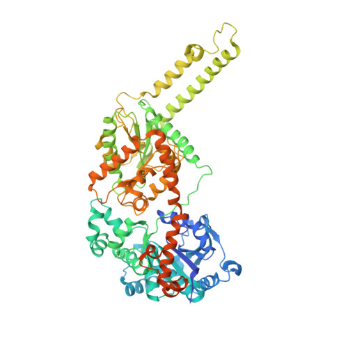
Structural basis for glucose-6-phosphate activation of glycogen synthase.

Baskaran, S.Roach, P.J.Depaoli-Roach, A.A.Hurley, T.D.

(2010) Proc Natl Acad Sci U S A 107: 17563-17568

  • DOI: https://doi.org/10.1073/pnas.1006340107
  • Primary Citation Related Structures: 
    3NAZ, 3NB0, 3NCH, 3O3C

  • PubMed Abstract: 

    Regulation of the storage of glycogen, one of the major energy reserves, is of utmost metabolic importance. In eukaryotes, this regulation is accomplished through glucose-6-phosphate levels and protein phosphorylation. Glycogen synthase homologs in bacteria and archaea lack regulation, while the eukaryotic enzymes are inhibited by protein kinase mediated phosphorylation and activated by protein phosphatases and glucose-6-phosphate binding. We determined the crystal structures corresponding to the basal activity state and glucose-6-phosphate activated state of yeast glycogen synthase-2. The enzyme is assembled into an unusual tetramer by an insertion unique to the eukaryotic enzymes, and this subunit interface is rearranged by the binding of glucose-6-phosphate, which frees the active site cleft and facilitates catalysis. Using both mutagenesis and intein-mediated phospho-peptide ligation experiments, we demonstrate that the enzyme's response to glucose-6-phosphate is controlled by Arg583 and Arg587, while four additional arginine residues present within the same regulatory helix regulate the response to phosphorylation.


  • Organizational Affiliation
    • Department of Biochemistry and Molelcular Biology, Indiana University School of Medicine, Indianapolis, IN 46202-5122, USA.

Macromolecule Content 

  • Total Structure Weight: 330.99 kDa 
  • Atom Count: 21,392 
  • Modeled Residue Count: 2,565 
  • Deposited Residue Count: 2,900 
  • Unique protein chains: 1

Macromolecules

Find similar proteins by:|  3D Structure
Entity ID: 1
MoleculeChains  Sequence LengthOrganismDetailsImage
Glycogen [starch] synthase isoform 2
A, B, C, D
725Saccharomyces cerevisiaeMutation(s): 2 
Gene Names: GSY2L8479.8YLR258W
EC: 2.4.1.11
UniProt
Find proteins for P27472 (Saccharomyces cerevisiae (strain ATCC 204508 / S288c))
Explore P27472 
Go to UniProtKB:  P27472
Entity Groups
Sequence Clusters30% Identity50% Identity70% Identity90% Identity95% Identity100% Identity
UniProt GroupP27472
Sequence Annotations
Expand
Reference Sequence

Small Molecules

Ligands 2 Unique
IDChains Name / Formula / InChI Key2D Diagram3D Interactions
G6P

Query on G6P


Download:Ideal Coordinates CCD File
E [auth A]
F [auth A]
I [auth B]
J [auth B]
M [auth C]
E [auth A],
F [auth A],
I [auth B],
J [auth B],
M [auth C],
O [auth D]
6-O-phosphono-alpha-D-glucopyranose
C6 H13 O9 P
NBSCHQHZLSJFNQ-DVKNGEFBSA-N
PEG

Query on PEG


Download:Ideal Coordinates CCD File
G [auth A]
H [auth B]
K [auth B]
L [auth B]
N [auth C]
G [auth A],
H [auth B],
K [auth B],
L [auth B],
N [auth C],
P [auth D]
DI(HYDROXYETHYL)ETHER
C4 H10 O3
MTHSVFCYNBDYFN-UHFFFAOYSA-N

Experimental Data & Validation

Experimental Data

  • Method: X-RAY DIFFRACTION
  • Resolution: 2.41 Å
  • R-Value Free:  0.242 (Depositor), 0.241 (DCC) 
  • R-Value Work:  0.211 (Depositor), 0.211 (DCC) 
  • R-Value Observed: 0.213 (Depositor) 
Space Group: I 2 2 2
Unit Cell:
Length ( Å )Angle ( ˚ )
a = 192.739α = 90
b = 206.982β = 90
c = 205.799γ = 90
Software Package:
Software NamePurpose
DENZOdata reduction
SCALEPACKdata scaling
PHENIXrefinement
PDB_EXTRACTdata extraction
HKL-2000data collection
HKL-2000data reduction
AMoREphasing

Structure Validation

View Full Validation Report


Ligand Structure Quality Assessment 


Entry History 

Deposition Data

Revision History  (Full details and data files)

  • Version 1.0: 2010-10-06
    Type: Initial release
  • Version 1.1: 2011-07-13
    Changes: Version format compliance
  • Version 1.2: 2017-11-08
    Changes: Refinement description
  • Version 1.3: 2020-07-29
    Type: Remediation
    Reason: Carbohydrate remediation
    Changes: Data collection, Database references, Derived calculations, Structure summary
  • Version 1.4: 2023-09-06
    Changes: Data collection, Database references, Refinement description, Structure summary