3M7H | pdb_00003m7h

Crystal structure of the bacteriocin LLPA from Pseudomonas sp.


Experimental Data Snapshot

  • Method: X-RAY DIFFRACTION
  • Resolution: 2.20 Å
  • R-Value Free: 
    0.204 (Depositor), 0.203 (DCC) 
  • R-Value Work: 
    0.172 (Depositor), 0.173 (DCC) 
  • R-Value Observed: 
    0.175 (Depositor) 

wwPDB Validation 3D Report Full Report

Validation slider image for 3M7H

This is version 1.4 of the entry. See complete history

Literature

Structural Determinants for Activity and Specificity of the Bacterial Toxin LlpA

Ghequire, M.G.Garcia-Pino, A.Lebbe, E.K.Spaepen, S.Loris, R.De Mot, R.

(2013) PLoS Pathog 9: e1003199-e1003199

  • DOI: https://doi.org/10.1371/journal.ppat.1003199
  • Primary Citation Related Structures: 
    3M7H, 3M7J, 4GC1, 4GC2

  • PubMed Abstract: 

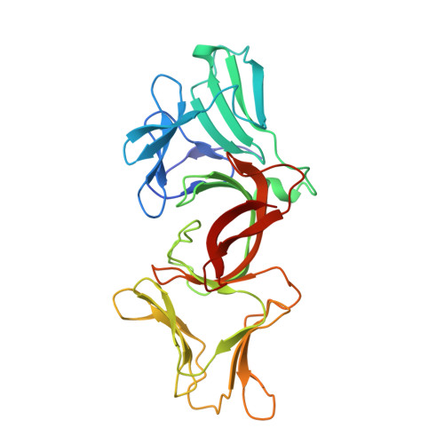
    Lectin-like bacteriotoxic proteins, identified in several plant-associated bacteria, are able to selectively kill closely related species, including several phytopathogens, such as Pseudomonas syringae and Xanthomonas species, but so far their mode of action remains unrevealed. The crystal structure of LlpABW, the prototype lectin-like bacteriocin from Pseudomonas putida, reveals an architecture of two monocot mannose-binding lectin (MMBL) domains and a C-terminal β-hairpin extension. The C-terminal MMBL domain (C-domain) adopts a fold very similar to MMBL domains from plant lectins and contains a binding site for mannose and oligomannosides. Mutational analysis indicates that an intact sugar-binding pocket in this domain is crucial for bactericidal activity. The N-terminal MMBL domain (N-domain) adopts the same fold but is structurally more divergent and lacks a functional mannose-binding site. Differential activity of engineered N/C-domain chimers derived from two LlpA homologues with different killing spectra, disclosed that the N-domain determines target specificity. Apparently this bacteriocin is assembled from two structurally similar domains that evolved separately towards dedicated functions in target recognition and bacteriotoxicity.


  • Organizational Affiliation
    • Centre of Microbial and Plant Genetics, University of Leuven, Heverlee-Leuven, Belgium.

Macromolecule Content 

  • Total Structure Weight: 59.99 kDa 
  • Atom Count: 4,225 
  • Modeled Residue Count: 542 
  • Deposited Residue Count: 552 
  • Unique protein chains: 1

Macromolecules

Find similar proteins by:|  3D Structure
Entity ID: 1
MoleculeChains  Sequence LengthOrganismDetailsImage
Putidacin L1
A, B
276Pseudomonas putidaMutation(s): 0 
Gene Names: llpA
UniProt
Find proteins for Q8GEJ9 (Pseudomonas putida)
Explore Q8GEJ9 
Go to UniProtKB:  Q8GEJ9
Entity Groups
Sequence Clusters30% Identity50% Identity70% Identity90% Identity95% Identity100% Identity
UniProt GroupQ8GEJ9
Sequence Annotations
Expand
Reference Sequence

Experimental Data & Validation

Experimental Data

  • Method: X-RAY DIFFRACTION
  • Resolution: 2.20 Å
  • R-Value Free:  0.204 (Depositor), 0.203 (DCC) 
  • R-Value Work:  0.172 (Depositor), 0.173 (DCC) 
  • R-Value Observed: 0.175 (Depositor) 
Space Group: P 21 21 2
Unit Cell:
Length ( Å )Angle ( ˚ )
a = 150.495α = 90
b = 154.492β = 90
c = 34.218γ = 90
Software Package:
Software NamePurpose
MAR345data collection
SHARPphasing
PHENIXrefinement
DENZOdata reduction
SCALEPACKdata scaling

Structure Validation

View Full Validation Report


Entry History 

Deposition Data

Revision History  (Full details and data files)

  • Version 1.0: 2011-03-23
    Type: Initial release
  • Version 1.1: 2011-07-13
    Changes: Version format compliance
  • Version 1.2: 2013-04-10
    Changes: Database references
  • Version 1.3: 2017-11-08
    Changes: Refinement description
  • Version 1.4: 2024-03-20
    Changes: Data collection, Database references