3LIZ | pdb_00003liz

crystal structure of bla g 2 complexed with Fab 4C3


Experimental Data Snapshot

  • Method: X-RAY DIFFRACTION
  • Resolution: 1.80 Å
  • R-Value Free: 
    0.202 (Depositor), 0.209 (DCC) 
  • R-Value Work: 
    0.177 (Depositor), 0.183 (DCC) 
  • R-Value Observed: 
    0.178 (Depositor) 

Starting Model: experimental
View more details

wwPDB Validation 3D Report Full Report

Validation slider image for 3LIZ

Ligand Structure Quality Assessment 


This is version 2.3 of the entry. See complete history

Literature

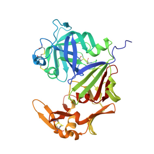
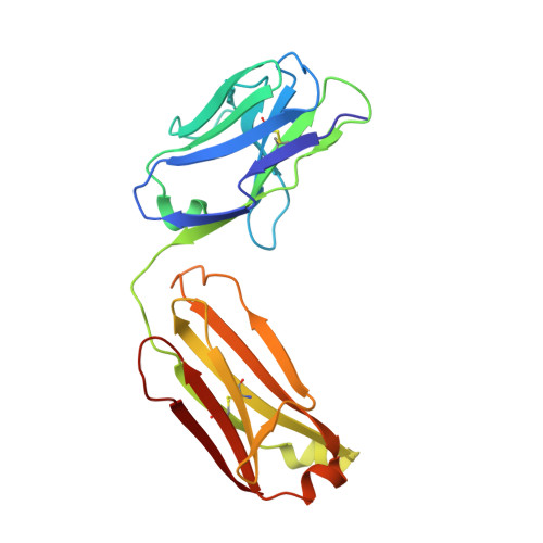
Mechanisms of allergen-antibody interaction of cockroach allergen Bla g 2 with monoclonal antibodies that inhibit IgE antibody binding.

Glesner, J.Wunschmann, S.Li, M.Gustchina, A.Wlodawer, A.Himly, M.Chapman, M.D.Pomes, A.

(2011) PLoS One 6: e22223-e22223

  • DOI: https://doi.org/10.1371/journal.pone.0022223
  • Primary Citation Related Structures: 
    3LIZ

  • PubMed Abstract: 

    Cockroach allergy is strongly associated with asthma, and involves the production of IgE antibodies against inhaled allergens. Reports of conformational epitopes on inhaled allergens are limited. The conformational epitopes for two specific monoclonal antibodies (mAb) that interfere with IgE antibody binding were identified by X-ray crystallography on opposite sites of the quasi-symmetrical cockroach allergen Bla g 2. Mutational analysis of selected residues in both epitopes was performed based on the X-ray crystal structures of the allergen with mAb Fab/Fab' fragments, to investigate the structural basis of allergen-antibody interactions. The epitopes of Bla g 2 for the mAb 7C11 or 4C3 were mutated, and the mutants were analyzed by SDS-PAGE, circular dichroism, and/or mass spectrometry. Mutants were tested for mAb and IgE antibody binding by ELISA and fluorescent multiplex array. Single or multiple mutations of five residues from both epitopes resulted in almost complete loss of mAb binding, without affecting the overall folding of the allergen. Preventing glycosylation by mutation N268Q reduced IgE binding, indicating a role of carbohydrates in the interaction. Cation-π interactions, as well as electrostatic and hydrophobic interactions, were important for mAb and IgE antibody binding. Quantitative differences in the effects of mutations on IgE antibody binding were observed, suggesting heterogeneity in epitope recognition among cockroach allergic patients. Analysis by site-directed mutagenesis of epitopes identified by X-ray crystallography revealed an overlap between monoclonal and IgE antibody binding sites and provided insight into the B cell repertoire to Bla g 2 and the mechanisms of allergen-antibody recognition, including involvement of carbohydrates.


  • Organizational Affiliation
    • INDOOR Biotechnologies, Inc, Charlottesville, Virginia, United States of America.

Macromolecule Content 

  • Total Structure Weight: 89.18 kDa 
  • Atom Count: 6,859 
  • Modeled Residue Count: 753 
  • Deposited Residue Count: 798 
  • Unique protein chains: 3

Macromolecules

Find similar proteins by:|  3D Structure
Entity ID: 1
MoleculeChains  Sequence LengthOrganismDetailsImage
Aspartic protease Bla g 2334Blattella germanicaMutation(s): 1 
EC: 3.4.23
UniProt
Find proteins for P54958 (Blattella germanica)
Explore P54958 
Go to UniProtKB:  P54958
Entity Groups
Sequence Clusters30% Identity50% Identity70% Identity90% Identity95% Identity100% Identity
UniProt GroupP54958
Glycosylation
Glycosylation Sites: 2
Sequence Annotations
Expand
Reference Sequence
Find similar proteins by:|  3D Structure
Entity ID: 2
MoleculeChains  Sequence LengthOrganismDetailsImage
4C3 monoclonal antibody Light ChainB [auth L]211Mus musculusMutation(s): 0 
Find similar proteins by:|  3D Structure
Entity ID: 3
MoleculeChains  Sequence LengthOrganismDetailsImage
4C3 monoclonal antibody Heavy ChainC [auth H]253Mus musculusMutation(s): 0 

Oligosaccharides

Help  
Entity ID: 4
MoleculeChains Length2D Diagram GlycosylationD Interactions
alpha-D-mannopyranose-(1-6)-beta-D-mannopyranose-(1-4)-2-acetamido-2-deoxy-beta-D-glucopyranose-(1-4)-2-acetamido-2-deoxy-beta-D-glucopyranoseD [auth B]4N-Glycosylation
Glycosylation Resources
GlyTouCan: G22573RC
GlyCosmos: G22573RC
GlyGen: G22573RC

Small Molecules

Ligands 4 Unique
IDChains Name / Formula / InChI Key2D Diagram3D Interactions
NAG

Query on NAG


Download:Ideal Coordinates CCD File
E [auth A]2-acetamido-2-deoxy-beta-D-glucopyranose
C8 H15 N O6
OVRNDRQMDRJTHS-FMDGEEDCSA-N
CD

Query on CD


Download:Ideal Coordinates CCD File
F [auth A],
G [auth A],
H [auth A]
CADMIUM ION
Cd
WLZRMCYVCSSEQC-UHFFFAOYSA-N
ZN

Query on ZN


Download:Ideal Coordinates CCD File
I [auth A]
J [auth A]
K [auth A]
L [auth A]
M [auth A]
I [auth A],
J [auth A],
K [auth A],
L [auth A],
M [auth A],
T [auth L],
U [auth L]
ZINC ION
Zn
PTFCDOFLOPIGGS-UHFFFAOYSA-N
EDO

Query on EDO


Download:Ideal Coordinates CCD File
N [auth A]
O [auth A]
P [auth A]
Q [auth A]
R [auth A]
N [auth A],
O [auth A],
P [auth A],
Q [auth A],
R [auth A],
S [auth A]
1,2-ETHANEDIOL
C2 H6 O2
LYCAIKOWRPUZTN-UHFFFAOYSA-N

Experimental Data & Validation

Experimental Data

  • Method: X-RAY DIFFRACTION
  • Resolution: 1.80 Å
  • R-Value Free:  0.202 (Depositor), 0.209 (DCC) 
  • R-Value Work:  0.177 (Depositor), 0.183 (DCC) 
  • R-Value Observed: 0.178 (Depositor) 
Space Group: C 1 2 1
Unit Cell:
Length ( Å )Angle ( ˚ )
a = 155.213α = 90
b = 105.285β = 132.58
c = 109.154γ = 90
Software Package:
Software NamePurpose
MAR345data collection
PHASERphasing
REFMACrefinement
HKL-3000data reduction
SCALEPACKdata scaling

Structure Validation

View Full Validation Report


Ligand Structure Quality Assessment 


Entry History 

Deposition Data

Revision History  (Full details and data files)

  • Version 1.0: 2010-12-15
    Type: Initial release
  • Version 1.1: 2011-07-13
    Changes: Version format compliance
  • Version 1.2: 2014-11-26
    Changes: Database references
  • Version 1.3: 2017-11-01
    Changes: Refinement description
  • Version 2.0: 2020-07-29
    Type: Remediation
    Reason: Carbohydrate remediation
    Changes: Atomic model, Data collection, Derived calculations, Structure summary
  • Version 2.1: 2021-10-13
    Changes: Database references, Structure summary
  • Version 2.2: 2023-09-06
    Changes: Data collection, Refinement description
  • Version 2.3: 2024-11-27
    Changes: Structure summary