3KNA

M296I mutant of foot-and-mouth disease virus RNA-polymerase in complex with a template- primer RNA


Experimental Data Snapshot

  • Method: X-RAY DIFFRACTION
  • Resolution: 2.80 Å
  • R-Value Free: 0.280 
  • R-Value Work: 0.239 
  • R-Value Observed: 0.241 

Starting Model: experimental
View more details

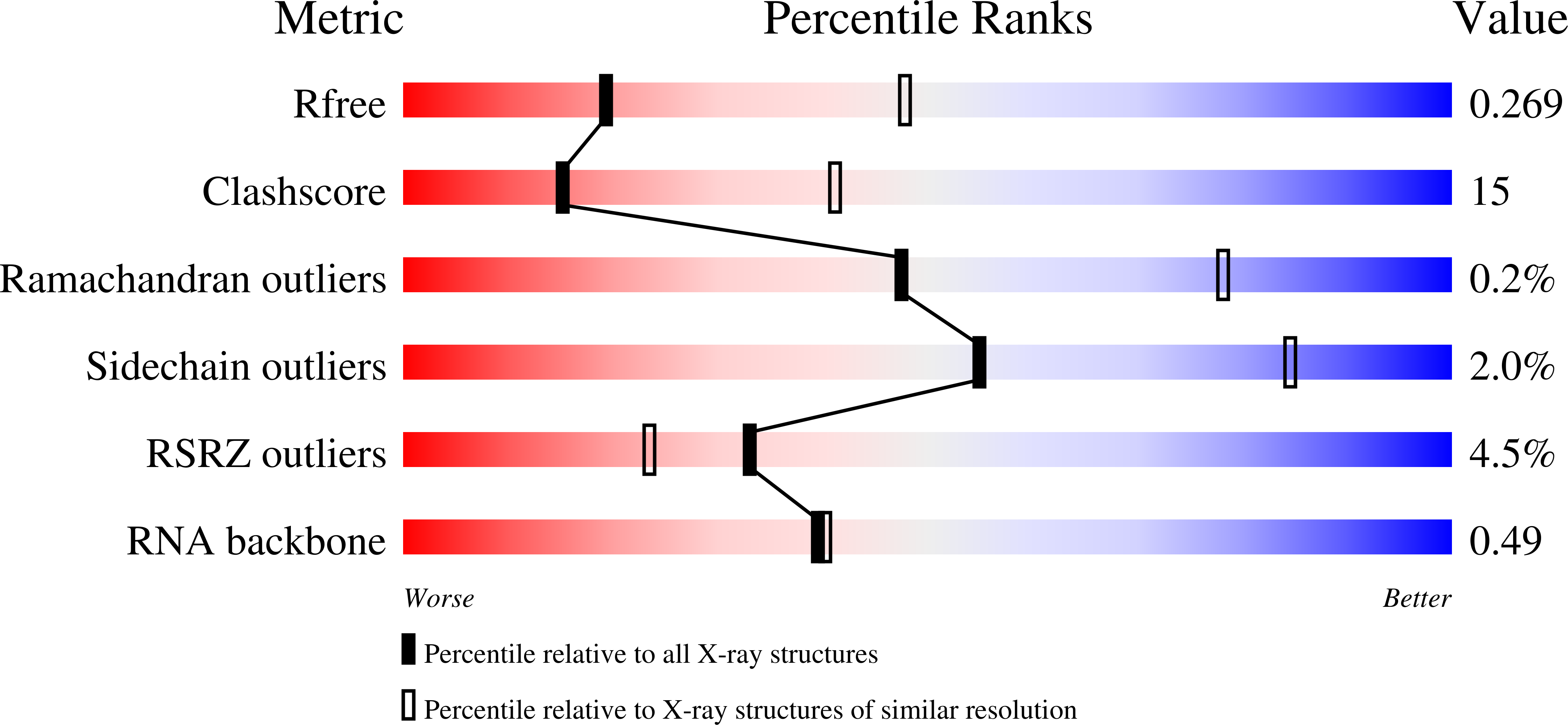
wwPDB Validation   3D Report Full Report


This is version 1.3 of the entry. See complete history


Literature

Structure of foot-and-mouth disease virus mutant polymerases with reduced sensitivity to ribavirin

Ferrer-Orta, C.Sierra, M.Agudo, R.de la Higuera, I.Arias, A.Perez-Luque, R.Escarmis, C.Domingo, E.Verdaguer, N.

(2010) J Virol 84: 6188-6199

  • DOI: https://doi.org/10.1128/JVI.02420-09
  • Primary Citation of Related Structures:  
    3KLV, 3KMQ, 3KMS, 3KNA, 3KOA

  • PubMed Abstract: 

    Passage of poliovirus (PV) or foot-and-mouth disease virus (FMDV) in the presence of ribavirin selected for viruses with decreased sensitivity to R, which included different mutations in their polymerase (3D): G64S located in the finger subdomain in the case of PV and M296I located within loop beta9-alpha11 at the active site in the case of FMDV. To investigate why disparate substitutions were selected in two closely related 3Ds, we constructed FMDVs with a 3D that included either G62S (the equivalent replacement in FMDV of PV G64S), M296I, or both substitutions. G62S, but not M296I, inflicts upon FMDV a strong selective disadvantage which is partially compensated for by the substitution M296I. The corresponding mutant polymerases, 3D(G62S), 3D(M296I), and 3D(G62S-M296I), were analyzed functionally and structurally. G62S in 3D impairs RNA-binding, polymerization, and R monophosphate incorporation activities. The X-ray structures of the 3D(G62S)-RNA, 3D(M296I)-RNA, and 3D(G62S-M296I)-RNA complexes show that although the two positions are separated by 13.1 A, the loops where the replacements reside are tightly connected through an extensive network of interactions that reach the polymerase active site. In particular, G62S seems to restrict the flexibility of loop beta9-alpha11 and, as a consequence, the flexibility of the active site and its ability to bind the RNA template. Thus, a localized change in the finger subdomain of 3D may affect the catalytic domain. The results provide a structural interpretation of why different amino acid substitutions were selected to confer R resistance in closely related viruses and reveal a complex network of intra-3D interactions that can affect the recognition of both the RNA template and incoming nucleotide.


  • Organizational Affiliation

    Institut de Biología Molecular de Barcelona (CSIC), Parc Cientific de Barcelona, Baldiri i Reixac 10, E-08028 Barcelona, Spain.


Macromolecules

Find similar proteins by:  (by identity cutoff)  |  3D Structure
Entity ID: 1
MoleculeChains Sequence LengthOrganismDetailsImage
3D polymerase476Foot and mouth disease virus CMutation(s): 1 
Gene Names: 3D
EC: 2.7.7.48
UniProt
Find proteins for Q9QCE3 (Foot and mouth disease virus C)
Explore Q9QCE3 
Go to UniProtKB:  Q9QCE3
Entity Groups  
Sequence Clusters30% Identity50% Identity70% Identity90% Identity95% Identity100% Identity
UniProt GroupQ9QCE3
Sequence Annotations
Expand
  • Reference Sequence

Find similar nucleic acids by:  Sequence   |   3D Structure  

Entity ID: 2
MoleculeChains LengthOrganismImage
RNA (5'-R(*AP*UP*GP*GP*GP*CP*C)-3')7N/A
Sequence Annotations
Expand
  • Reference Sequence

Find similar nucleic acids by:  Sequence   |   3D Structure  

Entity ID: 3
MoleculeChains LengthOrganismImage
RNA (5'-R(*GP*GP*CP*CP*C)-3')5N/A
Sequence Annotations
Expand
  • Reference Sequence
Small Molecules
Ligands 1 Unique
IDChains Name / Formula / InChI Key2D Diagram3D Interactions
MG
Query on MG

Download Ideal Coordinates CCD File 
D [auth A]MAGNESIUM ION
Mg
JLVVSXFLKOJNIY-UHFFFAOYSA-N
Experimental Data & Validation

Experimental Data

  • Method: X-RAY DIFFRACTION
  • Resolution: 2.80 Å
  • R-Value Free: 0.280 
  • R-Value Work: 0.239 
  • R-Value Observed: 0.241 
  • Space Group: P 41 21 2
Unit Cell:
Length ( Å )Angle ( ˚ )
a = 93.565α = 90
b = 93.565β = 90
c = 120.931γ = 90
Software Package:
Software NamePurpose
DNAdata collection
REFMACrefinement
MOSFLMdata reduction
SCALAdata scaling

Structure Validation

View Full Validation Report


Entry History 

Deposition Data

Revision History  (Full details and data files)

  • Version 1.0: 2010-07-07
    Type: Initial release
  • Version 1.1: 2011-07-13
    Changes: Version format compliance
  • Version 1.2: 2017-11-01
    Changes: Refinement description
  • Version 1.3: 2023-11-01
    Changes: Data collection, Database references, Derived calculations, Refinement description