3IIZ | pdb_00003iiz

X-ray structure of the FeFe-hydrogenase maturase HydE from T. maritima in complex with S-adenosyl-L-methionine


Experimental Data Snapshot

  • Method: X-RAY DIFFRACTION
  • Resolution: 1.62 Å
  • R-Value Free: 
    0.184 (Depositor), 0.188 (DCC) 
  • R-Value Work: 
    0.138 (Depositor), 0.148 (DCC) 
  • R-Value Observed: 
    0.140 (Depositor) 

Starting Model: experimental
View more details

wwPDB Validation 3D Report Full Report

Validation slider image for 3IIZ

Ligand Structure Quality Assessment 


This is version 1.4 of the entry. See complete history

Literature

Unexpected electron transfer mechanism upon AdoMet cleavage in radical SAM proteins

Nicolet, Y.Amara, P.Mouesca, J.-M.Fontecilla-Camps, J.C.

(2009) Proc Natl Acad Sci U S A 

  • DOI: https://doi.org/10.1073/pnas.0904385106
  • Primary Citation Related Structures: 
    3IIX, 3IIZ

  • PubMed Abstract: 

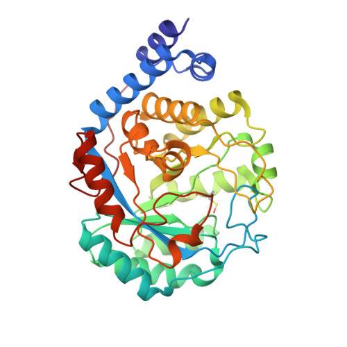
    Radical S-adenosine-L-methionine (SAM or AdoMet) proteins are involved in chemically difficult reactions including the synthesis of cofactors, the generation of protein radicals, and the maturation of complex organometallic catalytic sites. In the first and common step of the reaction, a conserved [Fe4S4] cluster donates an electron to perform the reductive cleavage of AdoMet into methionine and a reactive radical 5'-dA. species. The latter extracts a hydrogen atom from substrate eliciting one of the about 40 reactions so far characterized for this family of proteins. It has been suggested that the radical-generating mechanism differs depending on whether AdoMet is a cofactor or a substrate. It has also been speculated that electron transfer from the [Fe4S4] cluster to AdoMet is sulfur-based. Here we have used protein crystallography and theoretical calculations to show that regardless whether AdoMet serves as a cofactor or a substrate, the 5'-dA. generating mechanism should be common to the radical SAM proteins studied so far, and that electron transfer is mediated by a unique Fe from the conserved [Fe4S4] cluster. This unusual electron transfer is determined by the sulfonium ion in AdoMet.


  • Organizational Affiliation
    • Laboratoire de Cristallographie et Cristallogenèse des Protéines, Institut de Biologie Structurale J.P. Ebel, Commissariat à l'Energie Atomique, Centre National de la Recherche Scientifique, Université Joseph Fourier, 41 rue Jules Horowitz, 38027 Grenoble, France.

Macromolecule Content 

  • Total Structure Weight: 44.09 kDa 
  • Atom Count: 3,466 
  • Modeled Residue Count: 346 
  • Deposited Residue Count: 348 
  • Unique protein chains: 1

Macromolecules

Find similar proteins by:|  3D Structure
Entity ID: 1
MoleculeChains  Sequence LengthOrganismDetailsImage
Biotin synthetase, putative348Thermotoga maritimaMutation(s): 0 
Gene Names: TM_1269
EC: 1.8
UniProt
Find proteins for Q9X0Z6 (Thermotoga maritima (strain ATCC 43589 / DSM 3109 / JCM 10099 / NBRC 100826 / MSB8))
Explore Q9X0Z6 
Go to UniProtKB:  Q9X0Z6
Entity Groups
Sequence Clusters30% Identity50% Identity70% Identity90% Identity95% Identity100% Identity
UniProt GroupQ9X0Z6
Sequence Annotations
Expand
Reference Sequence

Small Molecules

Ligands 7 Unique
IDChains Name / Formula / InChI Key2D Diagram3D Interactions
CPS

Query on CPS


Download:Ideal Coordinates CCD File
D [auth A],
E [auth A],
F [auth A],
G [auth A],
H [auth A]
3-[(3-CHOLAMIDOPROPYL)DIMETHYLAMMONIO]-1-PROPANESULFONATE
C32 H58 N2 O7 S
UMCMPZBLKLEWAF-BCTGSCMUSA-N
SAM

Query on SAM


Download:Ideal Coordinates CCD File
C [auth A]S-ADENOSYLMETHIONINE
C15 H22 N6 O5 S
MEFKEPWMEQBLKI-FCKMPRQPSA-N
SF4

Query on SF4


Download:Ideal Coordinates CCD File
B [auth A]IRON/SULFUR CLUSTER
Fe4 S4
LJBDFODJNLIPKO-UHFFFAOYSA-N
FES

Query on FES


Download:Ideal Coordinates CCD File
I [auth A]FE2/S2 (INORGANIC) CLUSTER
Fe2 S2
NIXDOXVAJZFRNF-UHFFFAOYSA-N
CO3

Query on CO3


Download:Ideal Coordinates CCD File
N [auth A]CARBONATE ION
C O3
BVKZGUZCCUSVTD-UHFFFAOYSA-L
CL

Query on CL


Download:Ideal Coordinates CCD File
L [auth A],
M [auth A]
CHLORIDE ION
Cl
VEXZGXHMUGYJMC-UHFFFAOYSA-M
H2S

Query on H2S


Download:Ideal Coordinates CCD File
J [auth A],
K [auth A]
HYDROSULFURIC ACID
H2 S
RWSOTUBLDIXVET-UHFFFAOYSA-N
Modified Residues  1 Unique
IDChains TypeFormula2D DiagramParent
CSO
Query on CSO
A
L-PEPTIDE LINKINGC3 H7 N O3 SCYS
OTY
Query on OTY
A
L-PEPTIDE LINKINGC9 H11 N O4

--

Experimental Data & Validation

Experimental Data

  • Method: X-RAY DIFFRACTION
  • Resolution: 1.62 Å
  • R-Value Free:  0.184 (Depositor), 0.188 (DCC) 
  • R-Value Work:  0.138 (Depositor), 0.148 (DCC) 
  • R-Value Observed: 0.140 (Depositor) 
Space Group: P 21 21 21
Unit Cell:
Length ( Å )Angle ( ˚ )
a = 50.628α = 90
b = 78.78β = 90
c = 85.943γ = 90
Software Package:
Software NamePurpose
ProDCdata collection
REFMACrefinement
XDSdata reduction
XSCALEdata scaling
REFMACphasing

Structure Validation

View Full Validation Report


Ligand Structure Quality Assessment 


Entry History 

Deposition Data

Revision History  (Full details and data files)

  • Version 1.0: 2009-09-22
    Type: Initial release
  • Version 1.1: 2011-07-13
    Changes: Version format compliance
  • Version 1.2: 2017-11-01
    Changes: Refinement description
  • Version 1.3: 2023-11-01
    Changes: Data collection, Database references, Derived calculations, Refinement description
  • Version 1.4: 2023-11-22
    Changes: Data collection