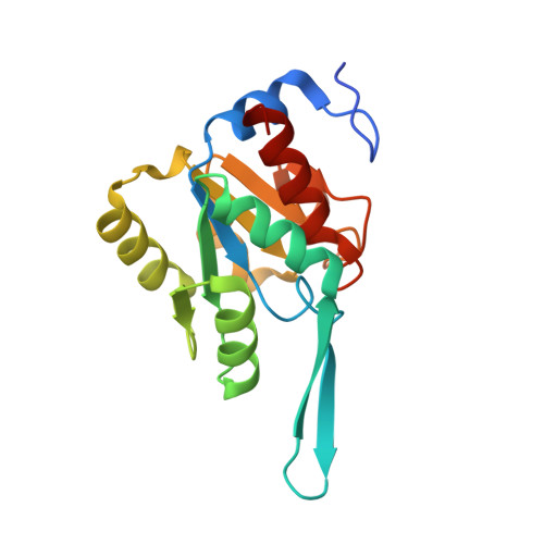
3I6B

Crystal structure of YrbI lacking the last 8 residues, in complex with Kdo and inorganic phosphate


Experimental Data Snapshot

  • Method: X-RAY DIFFRACTION
  • Resolution: 2.49 Å
  • R-Value Free: 0.238 
  • R-Value Work: 0.200 
  • R-Value Observed: 0.202 

Starting Model: experimental
View more details

wwPDB Validation   3D Report Full Report


Ligand Structure Quality Assessment 

Created with Raphaël 2.3.0Worse 01 BetterLigand structure goodness of fit to experimental dataBest fitted KDOClick on this verticalbar to view details

This is version 1.3 of the entry. See complete history


Literature

The Tail of KdsC: CONFORMATIONAL CHANGES CONTROL THE ACTIVITY OF A HALOACID DEHALOGENASE SUPERFAMILY PHOSPHATASE.

Biswas, T.Yi, L.Aggarwal, P.Wu, J.Rubin, J.R.Stuckey, J.A.Woodard, R.W.Tsodikov, O.V.

(2009) J Biol Chem 284: 30594-30603

  • DOI: https://doi.org/10.1074/jbc.M109.012278
  • Primary Citation of Related Structures:  
    2R8E, 2R8X, 2R8Y, 2R8Z, 3HYC, 3I6B

  • PubMed Abstract: 

    The phosphatase KdsC cleaves 3-deoxy-D-manno-octulosonate 8-phosphate to generate a molecule of inorganic phosphate and Kdo. Kdo is an essential component of the lipopolysaccharide envelope in Gram-negative bacteria. Because lipopolysaccharide is an important determinant of bacterial resistance and toxicity, KdsC is a potential target for novel antibacterial agents. KdsC belongs to the broad haloacid dehalogenase superfamily. In haloacid dehalogenase superfamily enzymes, substrate specificity and catalytic efficiency are generally dictated by a fold feature called the cap domain. It is therefore not clear why KdsC, which lacks a cap domain, is catalytically efficient and highly specific to 3-deoxy-D-manno-octulosonate 8-phosphate. Here, we present a set of seven structures of tetrameric Escherichia coli KdsC (ranging from 1.4 to 3.06 A in resolution) that model different intermediate states in its catalytic mechanism. A crystal structure of product-bound E. coli KdsC shows how the interface between adjacent monomers defines the active site pocket. Kdo is engaged in a network of polar and nonpolar interactions with residues at this interface, which explains substrate specificity. Furthermore, this structural and kinetic analysis strongly suggests that the binding of the flexible C-terminal region (tail) to the active site makes KdsC catalytically efficient by facilitating product release.


  • Organizational Affiliation

    Department of Medicinal Chemistry, College of Pharmacy, University of Michigan, Ann Arbor, Michigan 48109, USA.


Macromolecules
Find similar proteins by:  (by identity cutoff)  |  3D Structure
Entity ID: 1
MoleculeChains Sequence LengthOrganismDetailsImage
3-deoxy-D-manno-octulosonate 8-phosphate phosphatase
A, B, C, D
180Escherichia coli BMutation(s): 0 
Gene Names: c3958kdsCYrbI
EC: 3.1.3.45
UniProt
Find proteins for P67653 (Escherichia coli O6:H1 (strain CFT073 / ATCC 700928 / UPEC))
Explore P67653 
Go to UniProtKB:  P67653
Entity Groups  
Sequence Clusters30% Identity50% Identity70% Identity90% Identity95% Identity100% Identity
UniProt GroupP67653
Sequence Annotations
Expand
  • Reference Sequence
Small Molecules
Ligands 3 Unique
IDChains Name / Formula / InChI Key2D Diagram3D Interactions
KDO
Query on KDO

Download Ideal Coordinates CCD File 
I [auth B],
N [auth D]
3-deoxy-alpha-D-manno-oct-2-ulopyranosonic acid
C8 H14 O8
NNLZBVFSCVTSLA-HXUQBWEZSA-N
PO4
Query on PO4

Download Ideal Coordinates CCD File 
F [auth A],
H [auth B],
K [auth C],
M [auth D]
PHOSPHATE ION
O4 P
NBIIXXVUZAFLBC-UHFFFAOYSA-K
MG
Query on MG

Download Ideal Coordinates CCD File 
E [auth A],
G [auth B],
J [auth C],
L [auth D]
MAGNESIUM ION
Mg
JLVVSXFLKOJNIY-UHFFFAOYSA-N
Experimental Data & Validation

Experimental Data

  • Method: X-RAY DIFFRACTION
  • Resolution: 2.49 Å
  • R-Value Free: 0.238 
  • R-Value Work: 0.200 
  • R-Value Observed: 0.202 
  • Space Group: C 2 2 21
Unit Cell:
Length ( Å )Angle ( ˚ )
a = 64.964α = 90
b = 144.104β = 90
c = 145.854γ = 90
Software Package:
Software NamePurpose
HKL-2000data collection
PHASERphasing
REFMACrefinement
HKL-2000data reduction
HKL-2000data scaling

Structure Validation

View Full Validation Report


Ligand Structure Quality Assessment 

Created with Raphaël 2.3.0Worse 01 BetterLigand structure goodness of fit to experimental dataBest fitted KDOClick on this verticalbar to view details

Entry History 

Deposition Data

Revision History  (Full details and data files)

  • Version 1.0: 2009-09-01
    Type: Initial release
  • Version 1.1: 2011-07-13
    Changes: Advisory, Refinement description, Version format compliance
  • Version 1.2: 2020-07-29
    Type: Remediation
    Reason: Carbohydrate remediation
    Changes: Data collection, Derived calculations, Structure summary
  • Version 1.3: 2023-09-06
    Changes: Data collection, Database references, Refinement description, Structure summary