3H6Z | pdb_00003h6z

Crystal Structure of the Four MBT Repeats of Drosophila melanogaster Sfmbt in Complex with Peptide RHR (me)K VLR


Experimental Data Snapshot

  • Method: X-RAY DIFFRACTION
  • Resolution: 2.80 Å
  • R-Value Free: 
    0.254 (Depositor), 0.254 (DCC) 
  • R-Value Work: 
    0.221 (Depositor), 0.224 (DCC) 
  • R-Value Observed: 
    0.223 (Depositor) 

wwPDB Validation 3D Report Full Report

Validation slider image for 3H6Z

This is version 2.1 of the entry. See complete history

Literature

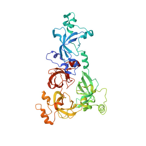
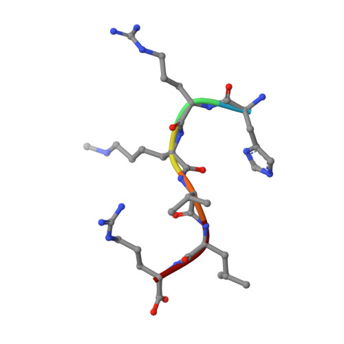
Molecular recognition of histone lysine methylation by the Polycomb group repressor dSfmbt

Grimm, C.Matos, R.Ly-Hartig, N.Steuerwald, U.Lindner, D.Rybin, V.Muller, J.Muller, C.W.

(2009) EMBO J 28: 1965-1977

  • DOI: https://doi.org/10.1038/emboj.2009.147
  • Primary Citation Related Structures: 
    3H6Z

  • PubMed Abstract: 

    Polycomb group (PcG) proteins repress transcription by modifying chromatin structure in target genes. dSfmbt is a subunit of the Drosophila melanogaster PcG protein complex PhoRC and contains four malignant brain tumour (MBT) repeats involved in the recognition of various mono- and dimethylated histone peptides. Here, we present the crystal structure of the four-MBT-repeat domain of dSfmbt in complex with a mono-methylated histone H4 peptide. Only a single histone peptide binds to the four-MBT-repeat domain. Mutational analyses show high-affinity binding with low peptide sequence selectivity through combinatorial interaction of the methyl-lysine with an aromatic cage and positively charged flanking residues with the surrounding negatively charged surface of the fourth MBT repeat. dSfmbt directly interacts with the PcG protein Scm, a related MBT-repeat protein with similar methyl-lysine binding activity. dSfmbt and Scm co-occupy Polycomb response elements of target genes in Drosophila and they strongly synergize in the repression of these target genes, suggesting that the combined action of these two MBT proteins is crucial for Polycomb silencing.


  • Organizational Affiliation
    • European Molecular Biology Laboratory, Grenoble Outstation, Grenoble, France.

Macromolecule Content 

  • Total Structure Weight: 103.75 kDa 
  • Atom Count: 7,115 
  • Modeled Residue Count: 874 
  • Deposited Residue Count: 906 
  • Unique protein chains: 2

Macromolecules

Find similar proteins by:|  3D Structure
Entity ID: 1
MoleculeChains  Sequence LengthOrganismDetailsImage
Polycomb protein SfmbtA,
C [auth B]
447Drosophila melanogasterMutation(s): 3 
Gene Names: Sfmbt
UniProt
Find proteins for Q9VK33 (Drosophila melanogaster)
Explore Q9VK33 
Go to UniProtKB:  Q9VK33
Entity Groups
Sequence Clusters30% Identity50% Identity70% Identity90% Identity95% Identity100% Identity
UniProt GroupQ9VK33
Sequence Annotations
Expand
Reference Sequence
Find similar proteins by:  Sequence   |   3D Structure  
Entity ID: 2
MoleculeChains  Sequence LengthOrganismDetailsImage
'HR(MLZ)VLRB [auth L],
D [auth M]
6synthetic constructMutation(s): 0 

Oligosaccharides

Help  
Entity ID: 3
MoleculeChains Length2D Diagram GlycosylationD Interactions
beta-D-fructofuranose-(2-1)-alpha-D-glucopyranoseE [auth C],
F [auth D],
G [auth E],
H [auth F]
2N/A
Glycosylation Resources
GlyTouCan: G05551OP
GlyCosmos: G05551OP

Small Molecules

Modified Residues  1 Unique
IDChains TypeFormula2D DiagramParent
MLZ
Query on MLZ
B [auth L],
D [auth M]
L-PEPTIDE LINKINGC7 H16 N2 O2LYS

Experimental Data & Validation

Experimental Data

  • Method: X-RAY DIFFRACTION
  • Resolution: 2.80 Å
  • R-Value Free:  0.254 (Depositor), 0.254 (DCC) 
  • R-Value Work:  0.221 (Depositor), 0.224 (DCC) 
  • R-Value Observed: 0.223 (Depositor) 
Space Group: P 2 21 21
Unit Cell:
Length ( Å )Angle ( ˚ )
a = 70.8α = 90
b = 97.04β = 90
c = 214.12γ = 90
Software Package:
Software NamePurpose
REFMACrefinement
PDB_EXTRACTdata extraction
ADSCdata collection
XDSdata reduction
XSCALEdata scaling
SHARPphasing

Structure Validation

View Full Validation Report


Entry History 

Deposition Data

Revision History  (Full details and data files)

  • Version 1.0: 2009-06-16
    Type: Initial release
  • Version 1.1: 2011-07-13
    Changes: Advisory, Refinement description, Version format compliance
  • Version 1.2: 2019-08-28
    Changes: Data collection, Derived calculations, Source and taxonomy
  • Version 1.3: 2019-10-16
    Changes: Data collection, Refinement description
  • Version 2.0: 2020-07-29
    Type: Remediation
    Reason: Carbohydrate remediation
    Changes: Atomic model, Data collection, Database references, Derived calculations, Non-polymer description, Structure summary
  • Version 2.1: 2021-11-10
    Changes: Database references, Structure summary