3H3W | pdb_00003h3w

Fitting of the gp6 crystal structure into 3D cryo-EM reconstruction of bacteriophage T4 dome-shaped baseplate


Experimental Data Snapshot

  • Method: ELECTRON MICROSCOPY
  • Resolution: 12.0 Å
  • Aggregation State: PARTICLE 
  • Reconstruction Method: SINGLE PARTICLE 

Starting Model: experimental
View more details

wwPDB Validation 3D Report Full Report

Validation slider image for 3H3W

This is version 1.3 of the entry. See complete history

Literature

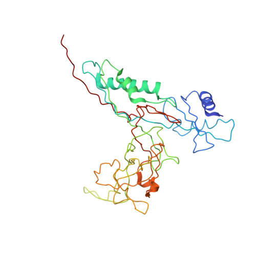
The structure of gene product 6 of bacteriophage T4, the hinge-pin of the baseplate.

Aksyuk, A.A.Leiman, P.G.Shneider, M.M.Mesyanzhinov, V.V.Rossmann, M.G.

(2009) Structure 17: 800-808

  • DOI: https://doi.org/10.1016/j.str.2009.04.005
  • Primary Citation Related Structures: 
    3H2T, 3H3W, 3H3Y

  • PubMed Abstract: 

    The baseplate of bacteriophage T4 is a multicomponent protein complex, which controls phage attachment to the host. It assembles from six wedges and a central hub. During infection the baseplate undergoes a large conformational change from a dome-shaped to a flat, star-shaped structure. We report the crystal structure of the C-terminal half of gene product (gp) 6 and investigate its motion with respect to the other proteins during the baseplate rearrangement. Six gp6 dimers interdigitate, forming a ring that maintains the integrity of the baseplate in both conformations. One baseplate wedge contains an N-terminal dimer of gp6, whereas neighboring wedges are tied together through the C-terminal dimer of gp6. The dimeric interactions are preserved throughout the rearrangement of the baseplate. However, the hinge angle between the N- and C-terminal parts of gp6 changes by approximately 15 degrees , accounting for a 10 A radial increase in the diameter of the gp6 ring.


  • Organizational Affiliation
    • Department of Biological Sciences, Purdue University, 915 W. State Street, West Lafayette, IN 47907-2054, USA.

Macromolecule Content 

  • Total Structure Weight: 459.65 kDa 
  • Atom Count: 31,380 
  • Modeled Residue Count: 3,900 
  • Deposited Residue Count: 4,020 
  • Unique protein chains: 1

Macromolecules

Find similar proteins by:|  3D Structure
Entity ID: 1
MoleculeChains  Sequence LengthOrganismDetailsImage
Baseplate structural protein Gp6
A, B, C, D, E
A, B, C, D, E, F, G, H, I, J, K, L
335Tequatrovirus T4Mutation(s): 0 
Gene Names: 6
UniProt
Find proteins for P19060 (Enterobacteria phage T4)
Explore P19060 
Go to UniProtKB:  P19060
Entity Groups
Sequence Clusters30% Identity50% Identity70% Identity90% Identity95% Identity100% Identity
UniProt GroupP19060
Sequence Annotations
Expand
Reference Sequence

Experimental Data & Validation

Experimental Data

  • Method: ELECTRON MICROSCOPY
  • Resolution: 12.0 Å
  • Aggregation State: PARTICLE 
  • Reconstruction Method: SINGLE PARTICLE 
EM Software:
TaskSoftware PackageVersion
RECONSTRUCTIONSPIDER

Structure Validation

View Full Validation Report


Entry History 

Deposition Data

Revision History  (Full details and data files)

  • Version 1.0: 2009-05-19
    Type: Initial release
  • Version 1.1: 2011-07-13
    Changes: Version format compliance
  • Version 1.2: 2018-07-18
    Changes: Data collection
  • Version 1.3: 2024-02-21
    Changes: Data collection, Database references, Refinement description