3H3R | pdb_00003h3r

Crystal structure of the CERT START domain in complex with HPA-14


Experimental Data Snapshot

  • Method: X-RAY DIFFRACTION
  • Resolution: 1.85 Å
  • R-Value Free: 
    0.253 (Depositor), 0.252 (DCC) 
  • R-Value Work: 
    0.203 (Depositor), 0.202 (DCC) 
  • R-Value Observed: 
    0.205 (Depositor) 

Starting Model: experimental
View more details

wwPDB Validation 3D Report Full Report

Validation slider image for 3H3R

Ligand Structure Quality Assessment 


This is version 1.3 of the entry. See complete history

Literature

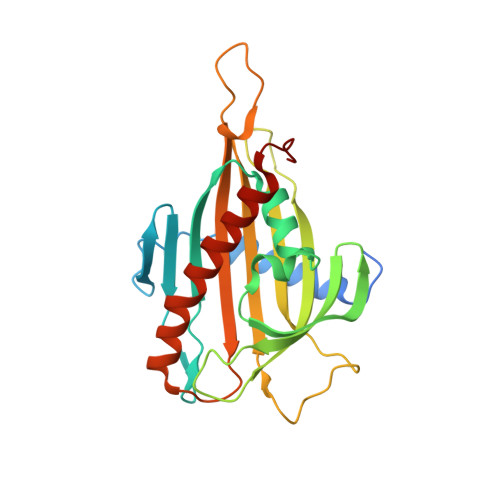
Crystal structures of the CERT START domain with inhibitors provide insights into the mechanism of ceramide transfer.

Kudo, N.Kumagai, K.Matsubara, R.Kobayashi, S.Hanada, K.Wakatsuki, S.Kato, R.

(2010) J Mol Biology 396: 245-251

  • DOI: https://doi.org/10.1016/j.jmb.2009.12.029
  • Primary Citation Related Structures: 
    3H3Q, 3H3R, 3H3S, 3H3T

  • PubMed Abstract: 

    The cytosolic protein CERT transfers ceramide from the endoplasmic reticulum to the Golgi apparatus where ceramide is converted to SM. The C-terminal START (steroidogenic acute regulatory protein-related lipid transfer) domain of CERT binds one ceramide molecule in its central amphiphilic cavity. (1R,3R)-N-(3-Hydroxy-1-hydroxymethyl-3-phenylpropyl)alkanamide (HPA), a synthesized analogue of ceramide, inhibits ceramide transfer by CERT. Here we report crystal structures of the CERT START domain in complex with HPAs of varying acyl chain lengths. In these structures, one HPA molecule is buried in the amphiphilic cavity where the amide and hydroxyl groups of HPA form a hydrogen-bond network with specific amino acid residues. The Omega1 loop, which has been suggested to function as a gate of the cavity, adopts a different conformation when bound to HPA than when bound to ceramide. In the Omega1 loop region, Trp473 shows the largest difference between these two structures. This residue exists inside of the cavity in HPA-bound structures, while it is exposed to the outside of the protein in the apo-form and ceramide-bound complex structures. Surface plasmon resonance experiments confirmed that Trp473 is important for interaction with membranes. These results provide insights into not only the molecular mechanism of inhibition by HPAs but also possible mechanisms by which CERT interacts with ceramide.


  • Organizational Affiliation
    • Structural Biology Research Center, Photon Factory, Institute of Materials Structure Science, High Energy Accelerator Research Organization (KEK), Tsukuba, Ibaraki, Japan.

Macromolecule Content 

  • Total Structure Weight: 58.65 kDa 
  • Atom Count: 4,016 
  • Modeled Residue Count: 471 
  • Deposited Residue Count: 510 
  • Unique protein chains: 1

Macromolecules

Find similar proteins by:|  3D Structure
Entity ID: 1
MoleculeChains  Sequence LengthOrganismDetailsImage
Goodpasture antigen binding protein
A, B
255Homo sapiensMutation(s): 0 
UniProt & NIH Common Fund Data Resources
Find proteins for Q9Y5P4 (Homo sapiens)
Explore Q9Y5P4 
Go to UniProtKB:  Q9Y5P4
PHAROS:  Q9Y5P4
GTEx:  ENSG00000113163 
Entity Groups
Sequence Clusters30% Identity50% Identity70% Identity90% Identity95% Identity100% Identity
UniProt GroupQ9Y5P4
Sequence Annotations
Expand
Reference Sequence

Small Molecules

Ligands 1 Unique
IDChains Name / Formula / InChI Key2D Diagram3D Interactions
14H

Query on 14H


Download:Ideal Coordinates CCD File
C [auth A],
D [auth B]
N-[(1R,3R)-3-hydroxy-1-(hydroxymethyl)-3-phenylpropyl]tetradecanamide
C24 H41 N O3
MWVNSXPNTJYJDR-DHIUTWEWSA-N

Experimental Data & Validation

Experimental Data

  • Method: X-RAY DIFFRACTION
  • Resolution: 1.85 Å
  • R-Value Free:  0.253 (Depositor), 0.252 (DCC) 
  • R-Value Work:  0.203 (Depositor), 0.202 (DCC) 
  • R-Value Observed: 0.205 (Depositor) 
Space Group: P 1 21 1
Unit Cell:
Length ( Å )Angle ( ˚ )
a = 42.417α = 90
b = 74.717β = 102.86
c = 77.713γ = 90
Software Package:
Software NamePurpose
DENZOdata reduction
SCALEPACKdata scaling
MOLREPphasing
REFMACrefinement
PDB_EXTRACTdata extraction
HKL-2000data collection
HKL-2000data reduction
HKL-2000data scaling

Structure Validation

View Full Validation Report


Ligand Structure Quality Assessment 


Entry History 

Deposition Data

Revision History  (Full details and data files)

  • Version 1.0: 2010-03-02
    Type: Initial release
  • Version 1.1: 2011-07-13
    Changes: Version format compliance
  • Version 1.2: 2017-11-01
    Changes: Refinement description
  • Version 1.3: 2023-11-01
    Changes: Data collection, Database references, Derived calculations, Refinement description