3GLB | pdb_00003glb

Crystal structure of the effector binding domain of a CATM variant (R156H)


Experimental Data Snapshot

  • Method: X-RAY DIFFRACTION
  • Resolution: 2.80 Å
  • R-Value Free: 
    0.279 (Depositor), 0.275 (DCC) 
  • R-Value Work: 
    0.205 (Depositor), 0.209 (DCC) 
  • R-Value Observed: 
    0.209 (Depositor) 

Starting Model: experimental
View more details

wwPDB Validation 3D Report Full Report

Validation slider image for 3GLB

This is version 1.4 of the entry. See complete history

Literature

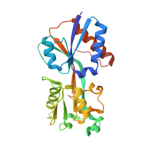
Inducer responses of BenM, a LysR-type transcriptional regulator from Acinetobacter baylyi ADP1.

Craven, S.H.Ezezika, O.C.Haddad, S.Hall, R.A.Momany, C.Neidle, E.L.

(2009) Mol Microbiol 72: 881-894

  • DOI: https://doi.org/10.1111/j.1365-2958.2009.06686.x
  • Primary Citation Related Structures: 
    2H99, 2H9B, 3GLB

  • PubMed Abstract: 

    BenM and CatM control transcription of a complex regulon for aromatic compound degradation. These Acinetobacter baylyi paralogues belong to the largest family of prokaryotic transcriptional regulators, the LysR-type proteins. Whereas BenM activates transcription synergistically in response to two effectors, benzoate and cis,cis-muconate, CatM responds only to cis,cis-muconate. Here, site-directed mutagenesis was used to determine the physiological significance of an unexpected benzoate-binding pocket in BenM discovered during structural studies. Residues in BenM were changed to match those of CatM in this hydrophobic pocket. Two BenM residues, R160 and Y293, were found to mediate the response to benzoate. Additionally, alteration of these residues caused benzoate to inhibit activation by cis,cis-muconate, positioned in a separate primary effector-binding site of BenM. The location of the primary site, in an interdomain cleft, is conserved in diverse LysR-type regulators. To improve understanding of this important family, additional regulatory mutants were analysed. The atomic-level structures were characterized of the effector-binding domains of variants that do not require inducers for activation, CatM(R156H) and BenM(R156H,T157S). These structures clearly resemble those of the wild-type proteins in their activated muconate-bound complexes. Amino acid replacements that enable activation without effectors reside at protein interfaces that may impact transcription through effects on oligomerization.


  • Organizational Affiliation
    • Department of Microbiology, University of Georgia, Athens, GA 30602-2605, USA.

Macromolecule Content 

  • Total Structure Weight: 102.66 kDa 
  • Atom Count: 7,099 
  • Modeled Residue Count: 858 
  • Deposited Residue Count: 900 
  • Unique protein chains: 1

Macromolecules

Find similar proteins by:|  3D Structure
Entity ID: 1
MoleculeChains  Sequence LengthOrganismDetailsImage
HTH-type transcriptional regulator catM
A, B, C, D
225Acinetobacter baylyi ADP1Mutation(s): 1 
Gene Names: catMcatRACIAD1445
UniProt
Find proteins for P07774 (Acinetobacter baylyi (strain ATCC 33305 / BD413 / ADP1))
Explore P07774 
Go to UniProtKB:  P07774
Entity Groups
Sequence Clusters30% Identity50% Identity70% Identity90% Identity95% Identity100% Identity
UniProt GroupP07774
Sequence Annotations
Expand
Reference Sequence

Small Molecules

Ligands 3 Unique
IDChains Name / Formula / InChI Key2D Diagram3D Interactions
CCU

Query on CCU


Download:Ideal Coordinates CCD File
E [auth A],
G [auth B],
L [auth C],
N [auth D]
(2Z,4Z)-HEXA-2,4-DIENEDIOIC ACID
C6 H6 O4
TXXHDPDFNKHHGW-CCAGOZQPSA-N
SO4

Query on SO4


Download:Ideal Coordinates CCD File
H [auth B],
I [auth B],
J [auth B]
SULFATE ION
O4 S
QAOWNCQODCNURD-UHFFFAOYSA-L
GOL

Query on GOL


Download:Ideal Coordinates CCD File
F [auth A],
K [auth B],
M [auth C],
O [auth D],
P [auth D]
GLYCEROL
C3 H8 O3
PEDCQBHIVMGVHV-UHFFFAOYSA-N

Experimental Data & Validation

Experimental Data

  • Method: X-RAY DIFFRACTION
  • Resolution: 2.80 Å
  • R-Value Free:  0.279 (Depositor), 0.275 (DCC) 
  • R-Value Work:  0.205 (Depositor), 0.209 (DCC) 
  • R-Value Observed: 0.209 (Depositor) 
Space Group: P 21 21 2
Unit Cell:
Length ( Å )Angle ( ˚ )
a = 110.689α = 90
b = 115.41β = 90
c = 76.068γ = 90
Software Package:
Software NamePurpose
DENZOdata reduction
SCALEPACKdata scaling
MOLREPphasing
REFMACrefinement
PDB_EXTRACTdata extraction
HKL-2000data collection
HKL-2000data scaling

Structure Validation

View Full Validation Report


Entry History 

Deposition Data

Revision History  (Full details and data files)

  • Version 1.0: 2009-05-19
    Type: Initial release
  • Version 1.1: 2011-07-13
    Changes: Advisory, Refinement description, Version format compliance
  • Version 1.2: 2017-11-01
    Changes: Refinement description
  • Version 1.3: 2021-10-20
    Changes: Database references, Derived calculations
  • Version 1.4: 2023-09-06
    Changes: Data collection, Refinement description