3GL2 | pdb_00003gl2

Crystal structure of dicamba monooxygenase bound to dicamba


Experimental Data Snapshot

  • Method: X-RAY DIFFRACTION
  • Resolution: 2.10 Å
  • R-Value Free: 
    0.247 (Depositor), 0.250 (DCC) 
  • R-Value Work: 
    0.190 (Depositor), 0.194 (DCC) 
  • R-Value Observed: 
    0.193 (Depositor) 

Starting Model: experimental
View more details

wwPDB Validation 3D Report Full Report

Validation slider image for 3GL2

Ligand Structure Quality Assessment 


This is version 1.2 of the entry. See complete history

Literature

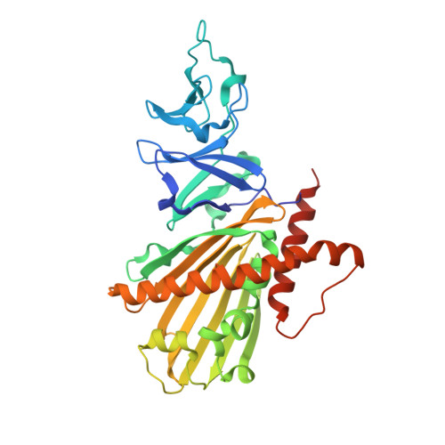
Crystal structure of dicamba monooxygenase: a Rieske nonheme oxygenase that catalyzes oxidative demethylation.

Dumitru, R.Jiang, W.Z.Weeks, D.P.Wilson, M.A.

(2009) J Mol Biology 392: 498-510

  • DOI: https://doi.org/10.1016/j.jmb.2009.07.021
  • Primary Citation Related Structures: 
    3GKE, 3GL0, 3GL2

  • PubMed Abstract: 

    Dicamba (3,6-dichloro-2-methoxybenzoic acid) is a widely used herbicide that is efficiently degraded by soil microbes. These microbes use a novel Rieske nonheme oxygenase, dicamba monooxygenase (DMO), to catalyze the oxidative demethylation of dicamba to 3,6-dichlorosalicylic acid (DCSA) and formaldehyde. We have determined the crystal structures of DMO in the free state, bound to its substrate dicamba, and bound to the product DCSA at 2.10-1.75 A resolution. The structures show that the DMO active site uses a combination of extensive hydrogen bonding and steric interactions to correctly orient chlorinated, ortho-substituted benzoic-acid-like substrates for catalysis. Unlike other Rieske aromatic oxygenases, DMO oxygenates the exocyclic methyl group, rather than the aromatic ring, of its substrate. This first crystal structure of a Rieske demethylase shows that the Rieske oxygenase structural scaffold can be co-opted to perform varied types of reactions on xenobiotic substrates.


  • Organizational Affiliation
    • Department of Biochemistry, The University of Nebraska-Lincoln, Lincoln, NE 68588-0664, USA.

Macromolecule Content 

  • Total Structure Weight: 117.01 kDa 
  • Atom Count: 8,156 
  • Modeled Residue Count: 989 
  • Deposited Residue Count: 1,047 
  • Unique protein chains: 1

Macromolecules

Find similar proteins by:|  3D Structure
Entity ID: 1
MoleculeChains  Sequence LengthOrganismDetailsImage
DdmC
A, B, C
349Stenotrophomonas maltophiliaMutation(s): 0 
Gene Names: ddmC
EC: 1.14.15
UniProt
Find proteins for Q5S3I3 (Stenotrophomonas maltophilia)
Explore Q5S3I3 
Go to UniProtKB:  Q5S3I3
Entity Groups
Sequence Clusters30% Identity50% Identity70% Identity90% Identity95% Identity100% Identity
UniProt GroupQ5S3I3
Sequence Annotations
Expand
Reference Sequence

Small Molecules

Ligands 3 Unique
IDChains Name / Formula / InChI Key2D Diagram3D Interactions
D3M

Query on D3M


Download:Ideal Coordinates CCD File
F [auth A],
I [auth B]
3,6-dichloro-2-methoxybenzoic acid
C8 H6 Cl2 O3
IWEDIXLBFLAXBO-UHFFFAOYSA-N
FES

Query on FES


Download:Ideal Coordinates CCD File
D [auth A],
G [auth B],
J [auth C]
FE2/S2 (INORGANIC) CLUSTER
Fe2 S2
NIXDOXVAJZFRNF-UHFFFAOYSA-N
FE

Query on FE


Download:Ideal Coordinates CCD File
E [auth A],
H [auth B]
FE (III) ION
Fe
VTLYFUHAOXGGBS-UHFFFAOYSA-N

Experimental Data & Validation

Experimental Data

  • Method: X-RAY DIFFRACTION
  • Resolution: 2.10 Å
  • R-Value Free:  0.247 (Depositor), 0.250 (DCC) 
  • R-Value Work:  0.190 (Depositor), 0.194 (DCC) 
  • R-Value Observed: 0.193 (Depositor) 
Space Group: P 32
Unit Cell:
Length ( Å )Angle ( ˚ )
a = 81.323α = 90
b = 81.323β = 90
c = 159.282γ = 120
Software Package:
Software NamePurpose
ADSCdata collection
PHASERphasing
REFMACrefinement
HKL-2000data reduction
HKL-2000data scaling

Structure Validation

View Full Validation Report


Ligand Structure Quality Assessment 


Entry History 

Deposition Data

Revision History  (Full details and data files)

  • Version 1.0: 2009-08-04
    Type: Initial release
  • Version 1.1: 2011-07-13
    Changes: Version format compliance
  • Version 1.2: 2023-09-06
    Changes: Data collection, Database references, Derived calculations, Refinement description