3G05 | pdb_00003g05

Crystal structure of N-terminal domain (2-550) of E.coli MnmG


Experimental Data Snapshot

  • Method: X-RAY DIFFRACTION
  • Resolution: 3.49 Å
  • R-Value Free: 
    0.265 (Depositor), 0.260 (DCC) 
  • R-Value Work: 
    0.227 (Depositor), 0.224 (DCC) 
  • R-Value Observed: 
    0.229 (Depositor) 

Starting Model: experimental
View more details

wwPDB Validation 3D Report Full Report

Validation slider image for 3G05

This is version 1.3 of the entry. See complete history

Literature

Structure-function analysis of Escherichia coli MnmG (GidA), a highly conserved tRNA-modifying enzyme.

Shi, R.Villarroya, M.Ruiz-Partida, R.Li, Y.Proteau, A.Prado, S.Moukadiri, I.Benitez-Paez, A.Lomas, R.Wagner, J.Matte, A.Velazquez-Campoy, A.Armengod, M.E.Cygler, M.

(2009) J Bacteriol 191: 7614-7619

  • DOI: https://doi.org/10.1128/JB.00650-09
  • Primary Citation Related Structures: 
    3CES, 3G05

  • PubMed Abstract: 

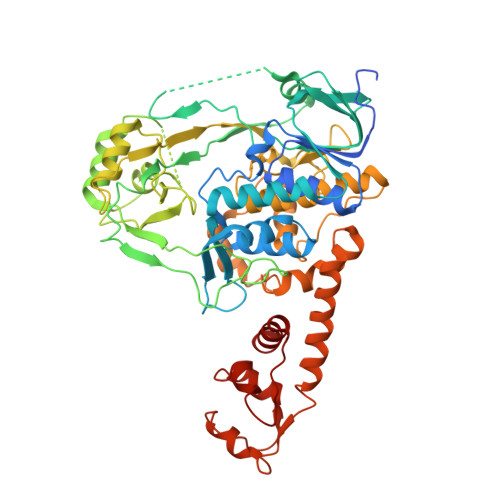
    The MnmE-MnmG complex is involved in tRNA modification. We have determined the crystal structure of Escherichia coli MnmG at 2.4-A resolution, mutated highly conserved residues with putative roles in flavin adenine dinucleotide (FAD) or tRNA binding and MnmE interaction, and analyzed the effects of these mutations in vivo and in vitro. Limited trypsinolysis of MnmG suggests significant conformational changes upon FAD binding.


  • Organizational Affiliation
    • Department of Biochemistry, McGill University, Montreal, Quebec, Canada.

Macromolecule Content 

  • Total Structure Weight: 128.28 kDa 
  • Atom Count: 8,156 
  • Modeled Residue Count: 1,051 
  • Deposited Residue Count: 1,152 
  • Unique protein chains: 1

Macromolecules

Find similar proteins by:|  3D Structure
Entity ID: 1
MoleculeChains  Sequence LengthOrganismDetailsImage
tRNA uridine 5-carboxymethylaminomethyl modification enzyme mnmG
A, B
576Escherichia coli O157:H7 str. EDL933Mutation(s): 0 
Gene Names: ECs4683gidAmnmGZ5241
UniProt
Find proteins for Q8XAY0 (Escherichia coli O157:H7)
Explore Q8XAY0 
Go to UniProtKB:  Q8XAY0
Entity Groups
Sequence Clusters30% Identity50% Identity70% Identity90% Identity95% Identity100% Identity
UniProt GroupQ8XAY0
Sequence Annotations
Expand
Reference Sequence

Experimental Data & Validation

Experimental Data

  • Method: X-RAY DIFFRACTION
  • Resolution: 3.49 Å
  • R-Value Free:  0.265 (Depositor), 0.260 (DCC) 
  • R-Value Work:  0.227 (Depositor), 0.224 (DCC) 
  • R-Value Observed: 0.229 (Depositor) 
Space Group: P 31 2 1
Unit Cell:
Length ( Å )Angle ( ˚ )
a = 144.593α = 90
b = 144.593β = 90
c = 271.019γ = 120
Software Package:
Software NamePurpose
DENZOdata reduction
SCALEPACKdata scaling
MOLREPphasing
REFMACrefinement
PDB_EXTRACTdata extraction
MAR345data collection

Structure Validation

View Full Validation Report


Entry History 

Deposition Data

Revision History  (Full details and data files)

  • Version 1.0: 2009-10-20
    Type: Initial release
  • Version 1.1: 2011-07-13
    Changes: Advisory, Refinement description, Version format compliance
  • Version 1.2: 2017-11-01
    Changes: Refinement description
  • Version 1.3: 2023-09-06
    Changes: Data collection, Database references, Derived calculations, Refinement description