3FZG | pdb_00003fzg

Structure of the 16S rRNA methylase ArmA


Experimental Data Snapshot

  • Method: X-RAY DIFFRACTION
  • Resolution: 2.00 Å
  • R-Value Free: 
    0.206 (Depositor), 0.205 (DCC) 
  • R-Value Work: 
    0.190 (Depositor), 0.186 (DCC) 
  • R-Value Observed: 
    0.190 (Depositor) 

wwPDB Validation 3D Report Full Report

Validation slider image for 3FZG

Ligand Structure Quality Assessment 


This is version 1.2 of the entry. See complete history

Literature

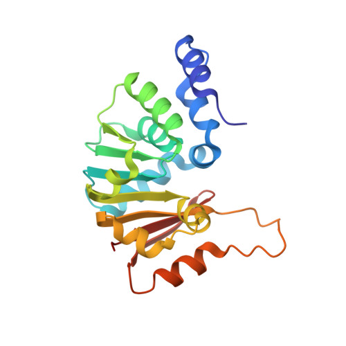
Structural bases for 16 S rRNA methylation catalyzed by ArmA and RmtB methyltransferases

Schmitt, E.Galimand, M.Panvert, M.Courvalin, P.Mechulam, Y.

(2009) J Mol Biology 388: 570-582

  • DOI: https://doi.org/10.1016/j.jmb.2009.03.034
  • Primary Citation Related Structures: 
    3FRH, 3FRI, 3FZG

  • PubMed Abstract: 

    Aminoglycosides are used extensively for the treatment of severe infections due to Gram-negative bacteria. However, certain species have become highly resistant after acquisition of genes for methyltransferases which catalyze post-transcriptional methylation of N7-G1405 in 16 S rRNA of 30 S ribosomal subunits. Inactivation of this enzymatic activity is therefore an important challenge for development of an effective therapy. The present work describes the crystallographic structures of methyltransferases RmtB and ArmA from clinical isolates. Together with biochemical experiments, the 3D structures indicate that the N-terminal domain specific for this family of methyltransferases is required for enzymatic activity. Site-directed mutagenesis has enabled important residues for catalysis and RNA binding to be identified. These high-resolution structures should underpin the design of potential inhibitors of these enzymes, which could be used to restore the activity of aminoglycosides against resistant pathogens.


  • Organizational Affiliation
    • Laboratoire de Biochimie, Ecole Polytechnique, Centre National de la Recherche Scientifique, Palaiseau Cedex, France. emma@botrytis.polytechnique.fr.

Macromolecule Content 

  • Total Structure Weight: 23.78 kDa 
  • Atom Count: 1,913 
  • Modeled Residue Count: 196 
  • Deposited Residue Count: 200 
  • Unique protein chains: 1

Macromolecules

Find similar proteins by:|  3D Structure
Entity ID: 1
MoleculeChains  Sequence LengthOrganismDetailsImage
16S rRNA methylase200Escherichia coliMutation(s): 0 
Gene Names: armApMUR050_016
EC: 2.1.1.179
UniProt
Find proteins for Q6F5A0 (Escherichia coli)
Explore Q6F5A0 
Go to UniProtKB:  Q6F5A0
Entity Groups
Sequence Clusters30% Identity50% Identity70% Identity90% Identity95% Identity100% Identity
UniProt GroupQ6F5A0
Sequence Annotations
Expand
Reference Sequence

Small Molecules

Ligands 1 Unique
IDChains Name / Formula / InChI Key2D Diagram3D Interactions
SAM

Query on SAM


Download:Ideal Coordinates CCD File
B [auth A]S-ADENOSYLMETHIONINE
C15 H22 N6 O5 S
MEFKEPWMEQBLKI-FCKMPRQPSA-N

Experimental Data & Validation

Experimental Data

  • Method: X-RAY DIFFRACTION
  • Resolution: 2.00 Å
  • R-Value Free:  0.206 (Depositor), 0.205 (DCC) 
  • R-Value Work:  0.190 (Depositor), 0.186 (DCC) 
  • R-Value Observed: 0.190 (Depositor) 
Space Group: C 2 2 21
Unit Cell:
Length ( Å )Angle ( ˚ )
a = 74.11α = 90
b = 90.08β = 90
c = 65.94γ = 90
Software Package:
Software NamePurpose
XDSdata scaling
SOLVEphasing
CNSrefinement
XDSdata reduction
XSCALEdata scaling

Structure Validation

View Full Validation Report


Ligand Structure Quality Assessment 


Entry History 

Deposition Data

Revision History  (Full details and data files)

  • Version 1.0: 2009-08-11
    Type: Initial release
  • Version 1.1: 2011-07-13
    Changes: Version format compliance
  • Version 1.2: 2024-03-20
    Changes: Data collection, Database references, Derived calculations