 Download Mendeley
SMY2-type GYF domain recognition in mRNA surveillance complexes
Ash, M.R., Faelber, K., Kosslick, D., Albert, G., Roske, Y., Kofler, M., Schuemann, M., Krause, E., Freund, C.To be published.
Experimental Data Snapshot
wwPDB Validation 3D Report Full Report

Entity ID: 1 | |||||
|---|---|---|---|---|---|
| Molecule | Chains | Sequence Length | Organism | Details | Image |
| Protein SMY2 | 100 | Saccharomyces cerevisiae | Mutation(s): 0 Gene Names: SMY2 | 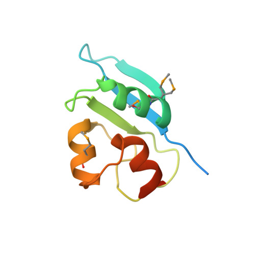 | |
UniProt | |||||
Entity Groups | |||||
| Sequence Clusters | 30% Identity50% Identity70% Identity90% Identity95% Identity100% Identity | ||||
| UniProt Group | P32909 | ||||
Sequence AnnotationsExpand | |||||
Reference Sequence | |||||
Entity ID: 2 | |||||
|---|---|---|---|---|---|
| Molecule | Chains | Sequence Length | Organism | Details | Image |
| Branchpoint-bridging protein | F [auth L], G [auth M], H [auth N], I [auth O], J [auth P] | 11 | N/A | Mutation(s): 0 | 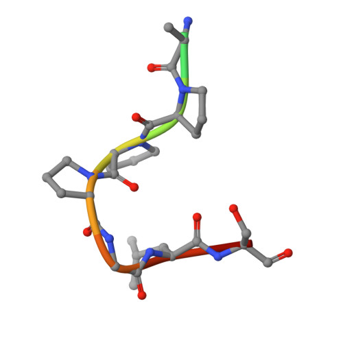 |
UniProt | |||||
Entity Groups | |||||
| Sequence Clusters | 30% Identity50% Identity70% Identity90% Identity95% Identity100% Identity | ||||
| UniProt Group | Q12186 | ||||
Sequence AnnotationsExpand | |||||
Reference Sequence | |||||
| Modified Residues 1 Unique | |||||
|---|---|---|---|---|---|
| ID | Chains | Type | Formula | 2D Diagram | Parent |
| MSE Query on MSE | A, B, C, D, E | L-PEPTIDE LINKING | C5 H11 N O2 Se |  | MET |
| Length ( Å ) | Angle ( ˚ ) |
|---|---|
| a = 101.4 | α = 90 |
| b = 101.4 | β = 90 |
| c = 150.7 | γ = 90 |
| Software Name | Purpose |
|---|---|
| MOSFLM | data reduction |
| SCALA | data scaling |
| REFMAC | refinement |
| PDB_EXTRACT | data extraction |
| SHELXCD | phasing |
| SHELXE | model building |
