3F5F | pdb_00003f5f

Crystal structure of heparan sulfate 2-O-sulfotransferase from gallus gallus as a maltose binding protein fusion.


Experimental Data Snapshot

  • Method: X-RAY DIFFRACTION
  • Resolution: 2.65 Å
  • R-Value Free: 
    0.243 (Depositor), 0.245 (DCC) 
  • R-Value Work: 
    0.200 (Depositor), 0.205 (DCC) 
  • R-Value Observed: 
    0.200 (Depositor) 

Starting Model: experimental
View more details

wwPDB Validation 3D Report Full Report

Validation slider image for 3F5F

Ligand Structure Quality Assessment 


This is version 2.3 of the entry. See complete history

Literature

Redirecting the substrate specificity of heparan sulfate 2-O-sulfotransferase by structurally guided mutagenesis.

Bethea, H.N.Xu, D.Liu, J.Pedersen, L.C.

(2008) Proc Natl Acad Sci U S A 105: 18724-18729

  • DOI: https://doi.org/10.1073/pnas.0806975105
  • Primary Citation Related Structures: 
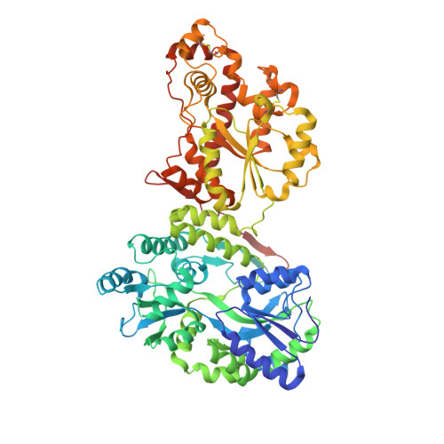
    3F5F

  • PubMed Abstract: 

    Heparan sulfate (HS) is a polysaccharide involved in essential physiological functions from regulating cell growth to blood coagulation. HS biosynthesis involves multiple specialized sulfotransferases such as 2-O-sulfotransferase (2OST) that transfers the sulfo group to the 2-OH position of iduronic acid (IdoA) or glucuronic acid (GlcA) within HS. Here, we report the homotrimeric crystal structure of 2OST from chicken, in complex with 3'-phosphoadenosine 5'-phosphate. Structural based mutational analysis has identified amino acid residues that are responsible for substrate specificity. The mutant R189A only transferred sulfates to GlcA moieties within the polysaccharide whereas mutants Y94A and H106A preferentially transferred sulfates to IdoA units. Our results demonstrate the feasibility for manipulating the substrate specificity of 2OST to synthesize HS with unique sulfation patterns. This work will aid the development of an enzymatic approach to synthesize heparin-based therapeutics.


  • Organizational Affiliation
    • Division of Medicinal Chemistry and Natural Products, Eshelman School of Pharmacy, University of North Carolina, Chapel Hill, NC 27599, USA.

Macromolecule Content 

  • Total Structure Weight: 75.06 kDa 
  • Atom Count: 5,403 
  • Modeled Residue Count: 656 
  • Deposited Residue Count: 658 
  • Unique protein chains: 1

Macromolecules

Find similar proteins by:|  3D Structure
Entity ID: 1
MoleculeChains  Sequence LengthOrganismDetailsImage
Maltose-binding periplasmic protein, Heparan sulfate 2-O-sulfotransferase 1658Escherichia coli K-12Gallus gallus
This entity is chimeric
Mutation(s): 1 
Gene Names: b4034HS2STHS2ST1JW3994malE
EC: 2.8.2
UniProt
Find proteins for P0AEX9 (Escherichia coli (strain K12))
Explore P0AEX9 
Go to UniProtKB:  P0AEX9
Find proteins for Q76KB1 (Gallus gallus)
Explore Q76KB1 
Go to UniProtKB:  Q76KB1
Entity Groups
Sequence Clusters30% Identity50% Identity70% Identity90% Identity95% Identity100% Identity
UniProt GroupsP0AEX9Q76KB1
Sequence Annotations
Expand
Reference Sequence

Oligosaccharides

Help  
Entity ID: 2
MoleculeChains Length2D Diagram GlycosylationD Interactions
alpha-D-glucopyranose-(1-4)-alpha-D-glucopyranose
B
2N/A
Glycosylation Resources
GlyTouCan: G07411ON
GlyCosmos: G07411ON

Small Molecules

Ligands 1 Unique
IDChains Name / Formula / InChI Key2D Diagram3D Interactions
A3P

Query on A3P


Download:Ideal Coordinates CCD File
C [auth A]ADENOSINE-3'-5'-DIPHOSPHATE
C10 H15 N5 O10 P2
WHTCPDAXWFLDIH-KQYNXXCUSA-N

Biologically Interesting Molecules (External Reference) 

1 Unique

Experimental Data & Validation

Experimental Data

  • Method: X-RAY DIFFRACTION
  • Resolution: 2.65 Å
  • R-Value Free:  0.243 (Depositor), 0.245 (DCC) 
  • R-Value Work:  0.200 (Depositor), 0.205 (DCC) 
  • R-Value Observed: 0.200 (Depositor) 
Space Group: P 63
Unit Cell:
Length ( Å )Angle ( ˚ )
a = 152.647α = 90
b = 152.647β = 90
c = 85.686γ = 120
Software Package:
Software NamePurpose
StructureStudiodata collection
MOLREPphasing
CNSrefinement
HKL-2000data reduction
HKL-2000data scaling

Structure Validation

View Full Validation Report


Ligand Structure Quality Assessment 


Entry History 

Deposition Data

Revision History  (Full details and data files)

  • Version 1.0: 2008-12-16
    Type: Initial release
  • Version 1.1: 2011-07-13
    Changes: Version format compliance
  • Version 1.2: 2017-08-23
    Changes: Refinement description, Source and taxonomy
  • Version 2.0: 2020-07-29
    Type: Remediation
    Reason: Carbohydrate remediation
    Changes: Atomic model, Data collection, Database references, Derived calculations, Structure summary
  • Version 2.1: 2021-10-20
    Changes: Database references, Structure summary
  • Version 2.2: 2023-09-06
    Changes: Data collection, Refinement description
  • Version 2.3: 2024-10-16
    Changes: Structure summary