3EUT | pdb_00003eut

X-ray crystal structure of a type III pentaketide synthase from Neurospora crassa


Experimental Data Snapshot

  • Method: X-RAY DIFFRACTION
  • Resolution: 2.00 Å
  • R-Value Free: 
    0.225 (Depositor), 0.228 (DCC) 
  • R-Value Work: 
    0.174 (Depositor), 0.176 (DCC) 
  • R-Value Observed: 
    0.176 (Depositor) 

wwPDB Validation 3D Report Full Report

Validation slider image for 3EUT

Ligand Structure Quality Assessment 


This is version 1.3 of the entry. See complete history

Literature

Distinct Structural Elements Dictate the Specificity of the Type III Pentaketide Synthase from Neurospora crassa.

Rubin-Pitel, S.B.Zhang, H.Vu, T.Brunzelle, J.S.Zhao, H.Nair, S.K.

(2008) Chem Biol 15: 1079-1090

  • DOI: https://doi.org/10.1016/j.chembiol.2008.08.011
  • Primary Citation Related Structures: 
    3EUO, 3EUQ, 3EUT

  • PubMed Abstract: 

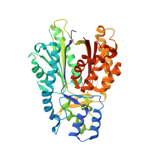
    The fungal type III polyketide synthase 2'-oxoalkylresorcylic acid synthase (ORAS) primes with a range of acyl-Coenzyme A thioesters (C4-C20) and extends using malonyl-Coenzyme A to produce pyrones, resorcinols, and resorcylic acids. To gain insight into this unusual substrate specificity and product profile, we have determined the crystal structures of ORAS to 1.75 A resolution, the Phe-252-->Gly site-directed mutant to 2.1 A resolution, and a binary complex of ORAS with eicosanoic acid to 2.0 A resolution. The structures reveal a distinct rearrangement of structural elements near the active site that allows accommodation of long-chain fatty acid esters and a reorientation of the gating mechanism that controls cyclization and polyketide chain length. The roles of these structural elements are further elucidated by characterization of various structure-based site-directed variants. These studies establish an unexpected plasticity to the PKS fold, unanticipated from structural studies of other members of this enzyme family.


  • Organizational Affiliation
    • Department of Chemical and Biomolecular Engineering, University of Illinois at Urbana-Champaign, 600 S. Mathews Avenue, Urbana, IL 61801, USA.

Macromolecule Content 

  • Total Structure Weight: 163.82 kDa 
  • Atom Count: 12,277 
  • Modeled Residue Count: 1,516 
  • Deposited Residue Count: 1,516 
  • Unique protein chains: 1

Macromolecules

Find similar proteins by:|  3D Structure
Entity ID: 1
MoleculeChains  Sequence LengthOrganismDetailsImage
Putative uncharacterized protein
A, B, C, D
379Neurospora crassaMutation(s): 0 
Gene Names: NCU04801NCU04801.1
UniProt
Find proteins for Q7S6N4 (Neurospora crassa (strain ATCC 24698 / 74-OR23-1A / CBS 708.71 / DSM 1257 / FGSC 987))
Explore Q7S6N4 
Go to UniProtKB:  Q7S6N4
Entity Groups
Sequence Clusters30% Identity50% Identity70% Identity90% Identity95% Identity100% Identity
UniProt GroupQ7S6N4
Sequence Annotations
Expand
Reference Sequence

Small Molecules

Modified Residues  1 Unique
IDChains TypeFormula2D DiagramParent
CSD
Query on CSD
A, B, C, D
L-PEPTIDE LINKINGC3 H7 N O4 SCYS

Experimental Data & Validation

Experimental Data

  • Method: X-RAY DIFFRACTION
  • Resolution: 2.00 Å
  • R-Value Free:  0.225 (Depositor), 0.228 (DCC) 
  • R-Value Work:  0.174 (Depositor), 0.176 (DCC) 
  • R-Value Observed: 0.176 (Depositor) 
Space Group: P 1 21 1
Unit Cell:
Length ( Å )Angle ( ˚ )
a = 68.867α = 90
b = 105.925β = 90.1
c = 104.124γ = 90
Software Package:
Software NamePurpose
REFMACrefinement

Structure Validation

View Full Validation Report


Ligand Structure Quality Assessment 


Entry History 

Deposition Data

Revision History  (Full details and data files)

  • Version 1.0: 2008-11-11
    Type: Initial release
  • Version 1.1: 2011-07-13
    Changes: Version format compliance
  • Version 1.2: 2023-12-27
    Changes: Data collection, Database references, Derived calculations
  • Version 1.3: 2024-11-20
    Changes: Structure summary