3DXA | pdb_00003dxa

Crystal Structure of the DM1 TCR in complex with HLA-B*4405 and decamer EBV antigen


Experimental Data Snapshot

  • Method: X-RAY DIFFRACTION
  • Resolution: 3.50 Å
  • R-Value Free: 
    0.330 (Depositor), 0.318 (DCC) 
  • R-Value Work: 
    0.286 (Depositor), 0.282 (DCC) 
  • R-Value Observed: 
    0.289 (Depositor) 

wwPDB Validation 3D Report Full Report

Validation slider image for 3DXA

This is version 1.3 of the entry. See complete history

Literature

Natural micropolymorphism in human leukocyte antigens provides a basis for genetic control of antigen recognition.

Archbold, J.K.Macdonald, W.A.Gras, S.Ely, L.K.Miles, J.J.Bell, M.J.Brennan, R.M.Beddoe, T.Wilce, M.C.Clements, C.S.Purcell, A.W.McCluskey, J.Burrows, S.R.Rossjohn, J.

(2009) J Exp Medicine 206: 209-219

  • DOI: https://doi.org/10.1084/jem.20082136
  • Primary Citation Related Structures: 
    3DX6, 3DX7, 3DX8, 3DX9, 3DXA

  • PubMed Abstract: 

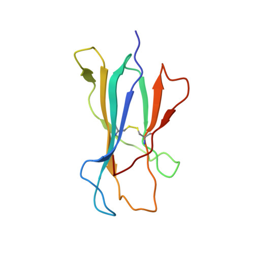
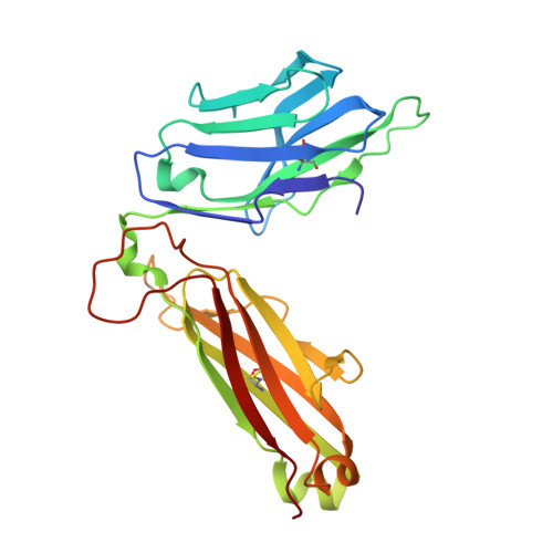
    Human leukocyte antigen (HLA) gene polymorphism plays a critical role in protective immunity, disease susceptibility, autoimmunity, and drug hypersensitivity, yet the basis of how HLA polymorphism influences T cell receptor (TCR) recognition is unclear. We examined how a natural micropolymorphism in HLA-B44, an important and large HLA allelic family, affected antigen recognition. T cell-mediated immunity to an Epstein-Barr virus determinant (EENLLDFVRF) is enhanced when HLA-B*4405 was the presenting allotype compared with HLA-B*4402 or HLA-B*4403, each of which differ by just one amino acid. The micropolymorphism in these HLA-B44 allotypes altered the mode of binding and dynamics of the bound viral epitope. The structure of the TCR-HLA-B*4405(EENLLDFVRF) complex revealed that peptide flexibility was a critical parameter in enabling preferential engagement with HLA-B*4405 in comparison to HLA-B*4402/03. Accordingly, major histocompatibility complex (MHC) polymorphism can alter the dynamics of the peptide-MHC landscape, resulting in fine-tuning of T cell responses between closely related allotypes.


  • Organizational Affiliation
    • The Protein Crystallography Unit, Department of Biochemistry and Molecular Biology, School of Biomedical Sciences, Monash University, Clayton, Victoria 3800, Australia.

Macromolecule Content 

  • Total Structure Weight: 286.36 kDa 
  • Atom Count: 20,171 
  • Modeled Residue Count: 2,484 
  • Deposited Residue Count: 2,484 
  • Unique protein chains: 5

Macromolecules

Find similar proteins by:|  3D Structure
Entity ID: 1
MoleculeChains  Sequence LengthOrganismDetailsImage
HLA class I histocompatibility complex HLA-B*4402
A, F, K
276Homo sapiensMutation(s): 1 
Gene Names: HLA-BHLAB
UniProt & NIH Common Fund Data Resources
Find proteins for P01889 (Homo sapiens)
Explore P01889 
Go to UniProtKB:  P01889
PHAROS:  P01889
GTEx:  ENSG00000234745 
Entity Groups
Sequence Clusters30% Identity50% Identity70% Identity90% Identity95% Identity100% Identity
UniProt GroupP01889
Sequence Annotations
Expand
Reference Sequence
Find similar proteins by:|  3D Structure
Entity ID: 2
MoleculeChains  Sequence LengthOrganismDetailsImage
Beta-2-microglobulin
B, G, L
99Homo sapiensMutation(s): 0 
Gene Names: B2MCDABP0092HDCMA22P
UniProt & NIH Common Fund Data Resources
Find proteins for P61769 (Homo sapiens)
Explore P61769 
Go to UniProtKB:  P61769
PHAROS:  P61769
GTEx:  ENSG00000166710 
Entity Groups
Sequence Clusters30% Identity50% Identity70% Identity90% Identity95% Identity100% Identity
UniProt GroupP61769
Sequence Annotations
Expand
Reference Sequence
Find similar proteins by:  Sequence   |   3D Structure  
Entity ID: 3
MoleculeChains  Sequence LengthOrganismDetailsImage
EBV decapeptide epitope
C, H, M
10N/AMutation(s): 0 
UniProt
Find proteins for P03204 (Epstein-Barr virus (strain B95-8))
Explore P03204 
Go to UniProtKB:  P03204
Entity Groups
Sequence Clusters30% Identity50% Identity70% Identity90% Identity95% Identity100% Identity
UniProt GroupP03204
Sequence Annotations
Expand
Reference Sequence
Find similar proteins by:|  3D Structure
Entity ID: 4
MoleculeChains  Sequence LengthOrganismDetailsImage
DM1 T cell receptor alpha chain
D, I, N
199Homo sapiensMutation(s): 0 
UniProt & NIH Common Fund Data Resources
Find proteins for P01848 (Homo sapiens)
Explore P01848 
Go to UniProtKB:  P01848
PHAROS:  P01848
Entity Groups
Sequence Clusters30% Identity50% Identity70% Identity90% Identity95% Identity100% Identity
UniProt GroupP01848
Sequence Annotations
Expand
Reference Sequence
Find similar proteins by:|  3D Structure
Entity ID: 5
MoleculeChains  Sequence LengthOrganismDetailsImage
DM1 T cell receptor beta chain
E, J, O
244Homo sapiensMutation(s): 0 
UniProt
Find proteins for P01850 (Homo sapiens)
Explore P01850 
Go to UniProtKB:  P01850
Entity Groups
Sequence Clusters30% Identity50% Identity70% Identity90% Identity95% Identity100% Identity
UniProt GroupP01850
Sequence Annotations
Expand
Reference Sequence

Experimental Data & Validation

Experimental Data

  • Method: X-RAY DIFFRACTION
  • Resolution: 3.50 Å
  • R-Value Free:  0.330 (Depositor), 0.318 (DCC) 
  • R-Value Work:  0.286 (Depositor), 0.282 (DCC) 
  • R-Value Observed: 0.289 (Depositor) 
Space Group: P 65 2 2
Unit Cell:
Length ( Å )Angle ( ˚ )
a = 121.954α = 90
b = 121.954β = 90
c = 695.914γ = 120
Software Package:
Software NamePurpose
XSCALEdata scaling
PHASERphasing
REFMACrefinement
PDB_EXTRACTdata extraction

Structure Validation

View Full Validation Report


Entry History 

Deposition Data

Revision History  (Full details and data files)

  • Version 1.0: 2009-01-27
    Type: Initial release
  • Version 1.1: 2011-07-13
    Changes: Advisory, Version format compliance
  • Version 1.2: 2021-10-20
    Changes: Database references
  • Version 1.3: 2024-11-13
    Changes: Data collection, Refinement description, Structure summary