3DFK | pdb_00003dfk

The crystal structure of teicoplanin pseudoaglycone deacetylase Orf2* bound to one of its products decanoic acid


Experimental Data Snapshot

  • Method: X-RAY DIFFRACTION
  • Resolution: 1.80 Å
  • R-Value Free: 
    0.220 (Depositor), 0.219 (DCC) 
  • R-Value Work: 
    0.176 (Depositor), 0.176 (DCC) 
  • R-Value Observed: 
    0.179 (Depositor) 

wwPDB Validation 3D Report Full Report

Validation slider image for 3DFK

Ligand Structure Quality Assessment 


This is version 1.2 of the entry. See complete history

Literature

Crystal structures of lipoglycopeptide antibiotic deacetylases: implications for the biosynthesis of a40926 and teicoplanin.

Zou, Y.Brunzelle, J.S.Nair, S.K.

(2008) Chem Biol 15: 533-545

  • DOI: https://doi.org/10.1016/j.chembiol.2008.05.009
  • Primary Citation Related Structures: 
    3DFF, 3DFI, 3DFK, 3DFM

  • PubMed Abstract: 

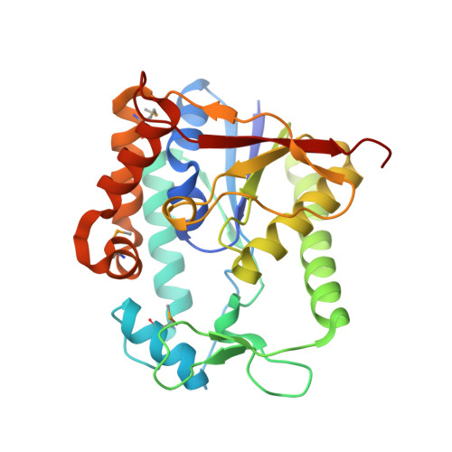
    The lipoglycopeptide antibiotics teicoplanin and A40926 have proven efficacy against Gram-positive pathogens. These drugs are distinguished from glycopeptide antibiotics by N-linked long chain acyl-D-glucosamine decorations that contribute to antibacterial efficacy. During the biosynthesis of lipoglycopeptides, tailoring glycosyltransferases attach an N-acetyl-D-glucosamine to the aglycone, and this N-acetyl-glucosaminyl pseudoaglycone is deacetylated prior to long chain hydrocarbon attachment. Here we present several high-resolution crystal structures of the pseudoaglycone deacetylases from the biosynthetic pathways of teicoplanin and A40926. The cocrystal structure of the teicoplanin pseudoaglycone deacetylase with a fatty acid product provides further insights into the roles of active-site residues, and suggests mechanistic similarities with structurally distinct zinc deacetylases, such as peptidoglycan deacetylase and LpxC. A unique, structurally mobile capping lid, located at the apex of these pseudoaglycone deacetylases, likely serves as a determinant of substrate specificity.


  • Organizational Affiliation
    • Department of Biochemistry, University of Illinois at Urbana-Champaign, Urbana, IL 61801, USA.

Macromolecule Content 

  • Total Structure Weight: 30.53 kDa 
  • Atom Count: 2,338 
  • Modeled Residue Count: 266 
  • Deposited Residue Count: 273 
  • Unique protein chains: 1

Macromolecules

Find similar proteins by:|  3D Structure
Entity ID: 1
MoleculeChains  Sequence LengthOrganismDetailsImage
Teicoplanin pseudoaglycone deacetylase Orf2273Actinoplanes teichomyceticusMutation(s): 0 
Gene Names: tcp14
UniProt
Find proteins for Q6ZZJ1 (Actinoplanes teichomyceticus)
Explore Q6ZZJ1 
Go to UniProtKB:  Q6ZZJ1
Entity Groups
Sequence Clusters30% Identity50% Identity70% Identity90% Identity95% Identity100% Identity
UniProt GroupQ6ZZJ1
Sequence Annotations
Expand
Reference Sequence

Small Molecules

Modified Residues  1 Unique
IDChains TypeFormula2D DiagramParent
MSE
Query on MSE
A
L-PEPTIDE LINKINGC5 H11 N O2 SeMET

Experimental Data & Validation

Experimental Data

  • Method: X-RAY DIFFRACTION
  • Resolution: 1.80 Å
  • R-Value Free:  0.220 (Depositor), 0.219 (DCC) 
  • R-Value Work:  0.176 (Depositor), 0.176 (DCC) 
  • R-Value Observed: 0.179 (Depositor) 
Space Group: P 1 21 1
Unit Cell:
Length ( Å )Angle ( ˚ )
a = 41.53α = 90
b = 64.263β = 99.01
c = 50.375γ = 90
Software Package:
Software NamePurpose
REFMACrefinement

Structure Validation

View Full Validation Report


Ligand Structure Quality Assessment 


Entry History 

Deposition Data

Revision History  (Full details and data files)

  • Version 1.0: 2008-07-22
    Type: Initial release
  • Version 1.1: 2011-07-13
    Changes: Version format compliance
  • Version 1.2: 2024-11-13
    Changes: Data collection, Database references, Derived calculations, Structure summary